CRISPR-Cas13 RNA-Targeting Revolution: Mechanism, Applications, and Future of Diagnostics & Therapeutics
This comprehensive review for researchers and drug development professionals examines the CRISPR-Cas13 system, focusing on its unique RNA-targeting mechanism and controversial collateral cleavage activity.
CRISPR-Cas13 RNA-Targeting Revolution: Mechanism, Applications, and Future of Diagnostics & Therapeutics
Abstract
This comprehensive review for researchers and drug development professionals examines the CRISPR-Cas13 system, focusing on its unique RNA-targeting mechanism and controversial collateral cleavage activity. We explore the foundational biology distinguishing Cas13 from DNA-editing Cas9, detail cutting-edge methodologies for diagnostics (SHERLOCK, DETECTR) and potential therapeutics, address critical troubleshooting and specificity optimization challenges, and provide a comparative analysis with other RNA-targeting platforms. The article synthesizes current research to guide experimental design and evaluate the translational potential of Cas13-based technologies in biomedicine.
Decoding Cas13: The RNA-Guided Scissor and Its Promiscuous Cleavage Activity
The CRISPR-Cas adaptive immune systems in prokaryotes have been repurposed as revolutionary biotechnological tools. The discovery and application of the DNA-targeting Cas9 endonuclease marked a watershed moment for genome editing. However, the emergence of the Class 2, Type VI CRISPR-Cas13 family represents a profound paradigm shift, enabling programmable targeting of RNA in mammalian cells without altering the genome. This whitepaper frames its technical discussion within the context of a broader thesis on the CRISPR-Cas13 mechanism of RNA targeting and its defining feature: non-specific, trans-acting "collateral cleavage" of bystander RNAs. This collateral activity, while initially seen as an off-target effect for editing, is now being harnessed for sensitive diagnostic tools and is a critical consideration for therapeutic development. This guide details the core mechanisms, comparative biology, experimental methodologies, and key reagents underpinning this shift from DNA to RNA targeting.
Comparative Core Mechanism: Cas9 vs. Cas13
Cas9 (e.g., SpCas9): A DNA endonuclease guided by a single-guide RNA (sgRNA). It recognizes a protospacer adjacent motif (PAM) in the target DNA, unwinds the duplex, and allows the guide RNA spacer to form an R-loop with the complementary DNA strand. This activates its two nuclease domains (HNH and RuvC) to create a double-strand break (DSB).
Cas13 (e.g., Cas13a/d, Cas13b): An RNA-guided, RNA-targeting ribonuclease. It recognizes a protospacer flanking site (PFS) in the target single-stranded RNA (ssRNA). Upon target RNA binding and cleavage by its two higher eukaryotes and prokaryotes nucleotide-binding (HEPN) domains, Cas13 undergoes a conformational change that activates its non-specific collateral RNase activity, leading to degradation of any nearby non-targeted RNA molecules.
Table 1: Key Quantitative Comparison of Cas9 (SpCas9) and Cas13 (LwaCas13a/RfxCas13d) Properties
| Property | Cas9 (SpCas9) | Cas13 (LwaCas13a) | Cas13 (RfxCas13d) |
|---|---|---|---|
| Target Molecule | dsDNA | ssRNA | ssRNA |
| Guide RNA | ~100-nt sgRNA | ~66-nt crRNA | ~69-nt crRNA |
| Recognition Site | 5'-NGG-3' PAM (DNA) | 3' non-G PFS (RNA) | Minimal PFS preference |
| Catalytic Domains | HNH, RuvC (DSB) | 2 x HPN (ssRNA cleavage) | 2 x HPN (ssRNA cleavage) |
| Primary Cleavage | Sequence-specific, cis | Sequence-specific, cis | Sequence-specific, cis |
| Collateral Activity | None | Promiscuous ssRNA cleavage (trans) | Attenuated ssRNA cleavage (trans) |
| Size (aa) | 1368 | 968-1250 | ~930 |
| Key Applications | Gene knockout, knock-in | RNA knockdown, diagnostics (e.g., SHERLOCK), viral inhibition | Mammalian RNA knockdown, base editing (REPAIR) |
Diagram 1: Cas9 vs Cas13 Core Mechanism (760px max)
Detailed Experimental Protocols
Protocol 1: Validating Cas13 RNA Knockdown in Mammalian Cells Objective: To assess sequence-specific RNA knockdown efficiency and specificity of a Cas13 effector (e.g., RfxCas13d).
- Construct Design: Clone the gene for a nuclear localization signal (NLS)-tagged RfxCas13d and an expression cassette for its cognate crRNA (targeting a gene of interest, GOI) into a mammalian expression plasmid (e.g., under EF1α/U6 promoters).
- Controls: Include a non-targeting crRNA control and a catalytically dead mutant (dCas13) control.
- Cell Transfection: Seed HEK293T cells in a 24-well plate. At 70-80% confluency, transfect with 500 ng of plasmid DNA per well using a transfection reagent like Lipofectamine 3000.
- Harvesting: 48-72 hours post-transfection, lyse cells in TRIzol reagent for RNA isolation.
- Analysis:
- qRT-PCR: Synthesize cDNA. Perform quantitative PCR with TaqMan or SYBR Green probes specific for the GOI and housekeeping genes (e.g., GAPDH, ACTB). Calculate fold-change using the ΔΔCt method.
- RNA Sequencing: For a global profile, prepare libraries from total RNA and perform next-generation sequencing. Analyze differential gene expression and potential off-target effects.
Protocol 2: Detecting Cas13 Collateral Cleavage Activity In Vitro Objective: To visualize and quantify the promiscuous RNase activity of Cas13 (e.g., LwaCas13a) upon target activation.
- Protein Purification: Express and purify His-tagged LwaCas13a protein from E. coli.
- RNA Preparation: In vitro transcribe and purify:
- Target RNA: A ~1-kb RNA containing the target sequence.
- Reporter RNA: A short (~100-200 nt), unrelated, fluorescently labelled (e.g., FAM at 5' end, quencher at 3' end) or biotinylated RNA.
- Non-target Control RNA: An RNA without complementarity to the crRNA.
- Reaction Setup: In a 20 µL reaction buffer (20 mM HEPES pH 6.8, 50 mM KCl, 5 mM MgCl₂, 1 mM DTT):
- Experimental: 50 nM LwaCas13a, 50 nM crRNA, 5 nM target RNA, 50 nM reporter RNA.
- Controls: Omit target RNA; use non-target control RNA.
- Incubation & Detection: Incubate at 37°C. Monitor reporter cleavage in real-time via fluorescence increase (if quenched reporter) or at endpoint by running products on a denaturing urea-PAGE gel and imaging for fluorescence or via streptavidin-horseradish peroxidase (if biotinylated).
The Cas13 Signaling Pathway: Target Detection to Collateral Cleavage
Diagram 2: Cas13 Activation & Collateral Cleavage Pathway (760px)
The Scientist's Toolkit: Key Research Reagent Solutions
Table 2: Essential Materials for Cas13 Research
| Reagent / Material | Function / Explanation | Example Supplier/Part Number |
|---|---|---|
| RfxCas13d (CasRx) Expression Plasmid | Mammalian codon-optimized version of Ruminococcus flavefaciens Cas13d. Preferred for efficient, specific RNA knockdown in mammalian cells with minimal collateral effects. | Addgene #109049 (pXR001: EF1α-CasRx-2xNLS) |
| LwaCas13a Expression & Purification System | Leptotrichia wadei Cas13a. High collateral activity makes it ideal for in vitro diagnostics (SHERLOCK). Often used from E. coli expression kits. | Addgene #90091 (pC005-Cas13a), NEB HiScribe T7 Kit for crRNA |
| Catalytically Dead Mutant (dCas13) | Cas13 with point mutations (e.g., RfxCas13d: H798A, H983A) in HEPN domains. Serves as a critical negative control for RNA cleavage-independent effects. | Addgene #109050 (pXR002: EF1α-dCasRx-2xNLS) |
| crRNA Cloning Backbone (U6-sgRNA) | Plasmid for expressing crRNA under RNA Polymerase III (U6) promoter in mammalian cells. Contains scaffold sequence for specific Cas13 binding. | Addgene #109053 (pXR003: U6-crRNA) |
| Fluorescent Quenched Reporter RNA (FQ-reporter) | Short RNA oligonucleotide with a 5' fluorophore (FAM) and a 3' quencher (Iowa Black). Collateral cleavage separates the pair, generating fluorescence. Core component of SHERLOCK. | Integrated DNA Technologies (IDT), custom synthesis |
| Recombinant RNase Inhibitor (e.g., SUPERase-In) | Inhibits common RNases but not Cas13's HEPN activity. Essential for handling RNA samples in Cas13 assays to prevent degradation from background RNases. | Thermo Fisher Scientific, AM2694 |
| Nucleofection Kit for Primary Cells | For efficient delivery of Cas13 RNP (ribonucleoprotein) complexes or plasmids into hard-to-transfect cell types (e.g., neurons, T cells). | Lonza, various cell-type specific kits |
| Cell-Free Transcription-Translation Mix (TXTL) | Rapid in vitro expression of Cas13 protein and crRNA for prototyping and diagnostic assay development without purification steps. | Arbor Biosciences, myTXTL Sigma 70 Kit |
The discovery of CRISPR-Cas systems has revolutionized programmable nucleic acid targeting. While Cas9 and Cas12 target DNA, Cas13 targets RNA. A defining feature of Type VI CRISPR-Cas13 effectors is their capacity for trans or "collateral" cleavage of non-target RNA upon target recognition, a phenomenon central to both diagnostic applications and fundamental biology. This activity is intrinsically linked to the Higher Eukaryotes and Prokaryotes Nucleotide-binding (HEPN) domain, a conserved superfamily found in numerous RNases. This whitepaper provides an in-depth technical analysis of HEPN domain architecture, its catalytic mechanism in RNA cleavage, and its role within the CRISPR-Cas13 system. The content is framed within the broader thesis that understanding HEPN domain dynamics is critical for harnessing Cas13 for precise RNA manipulation and for developing next-generation RNA-targeting therapeutics and diagnostics.
Structural Anatomy of the HEPN Domain
HEPN domains are typically ~130 amino acids in length and adopt a ferredoxin-like fold composed of a central β-sheet flanked by α-helices. In Cas13 proteins, two HEPN domains form the active nuclease core. Key structural motifs include:
- The RNase Active Site: Formed at the interface of the two HEPN domains (often termed HEPN1 and HEPN2). Catalysis is mediated by conserved motifs: the RxxxxH motif (where 'x' is any amino acid), with the Arg (R) and His (H) residues being absolutely critical for activity.
- The Pre-crRNA and Target RNA Binding Channel: A positively charged groove guides the crRNA:target RNA duplex towards the catalytic pocket.
- Allosteric Regulation Sites: Conformational changes upon crRNA:target RNA binding, particularly in the Helical-1 and Helical-2 domains of Cas13, trigger the repositioning of the HEPN domains from an auto-inhibited state to an active conformation.
Diagram 1: Cas13a Activation and Collateral Cleavage Pathway
Catalytic Mechanism of RNA Cleavage by HEPN Domains
The HEPN domain-catalyzed RNA cleavage is a metal-ion independent hydrolysis reaction. The conserved Arg and His residues act as a general base and acid, respectively, to activate a water molecule for in-line nucleophilic attack on the scissile phosphate.
- Substrate Positioning: Single-stranded RNA (ssRNA) substrate binds in the catalytic pocket.
- General Base Activation: The conserved Histidine deprotonates a water molecule, generating a nucleophilic hydroxide ion.
- Nucleophilic Attack: The hydroxide ion attacks the phosphorus atom at the scissile phosphate (typically 3' of a uridine or adenine), forming a pentavalent transition state.
- Proton Donation & Cleavage: The conserved Arginine stabilizes the transition state. The protonated histidine (or a second water) donates a proton to the 2'-OH of the ribose, facilitating the cleavage of the 5'-O bond.
- Product Release: Cleavage results in a 2',3'-cyclic phosphate and a 5'-OH termini, a hallmark of metal-independent RNase activity.
Table 1: Conserved Catalytic Motifs in HEPN-Domain RNases
| Protein/System | Conserved Motif (HEPN1) | Conserved Motif (HEPN2) | Cleavage Products | Metal Requirement |
|---|---|---|---|---|
| Cas13a (LshC2c2) | R...H (RxxxxH) | R...H (RxxxxH) | 2',3'-cyclic phosphate, 5'-OH | Independent |
| Cas13b (PbuC2c2) | R...H (RxxxxH) | R...H (RxxxxH) | 2',3'-cyclic phosphate, 5'-OH | Independent |
| Cas13d (RfxCas13d) | R...H (RxxxxH) | R...H (RxxxxH) | 2',3'-cyclic phosphate, 5'-OH | Independent |
| RNase L | R...H (RxxxxH) | N/A (pseudokinase domain) | 2',3'-cyclic phosphate, 5'-OH | Independent |
Key Experimental Protocols for HEPN Domain Analysis
Protocol 1: In Vitro Collateral Cleavage Assay (Fluorometric)
- Purpose: Quantify Cas13 HEPN domain nuclease activity and collateral cleavage kinetics.
- Reagents: Purified Cas13 protein, crRNA, target RNA trigger, quenched fluorescent RNA reporter (e.g., FAM-UUUUUU-BHQ1), RNase-free buffer.
- Procedure:
- Assemble reaction: 50 nM Cas13:crRNA complex, 5 nM target RNA, 1 µM fluorescent reporter in 20 µL reaction buffer.
- Incubate at 37°C in a real-time PCR instrument or plate reader.
- Monitor fluorescence (Ex/Em: 485/535 nm for FAM) every minute for 60-120 minutes.
- Calculate initial reaction velocity (RFU/min) and time to threshold.
Protocol 2: Structural Analysis via Cryo-Electron Microscopy (Cryo-EM)
- Purpose: Determine high-resolution structures of Cas13 in pre- and post-activation states.
- Procedure:
- Sample Preparation: Purify Cas13:crRNA:target RNA complexes to homogeneity. Apply 3-4 µL to a glow-discharged cryo-EM grid, blot, and plunge-freeze in liquid ethane.
- Data Collection: Collect multi-frame movies on a 300 keV cryo-TEM with a K3 direct electron detector at a nominal magnification of 81,000x (yielding ~1.0 Å/pixel).
- Image Processing: Motion correction, CTF estimation, particle picking (e.g., crYOLO), 2D classification, ab-initio reconstruction, and heterogeneous refinement in Relion or CryoSPARC to separate conformational states.
- Model Building: Fit existing atomic models into the map using Chimera/X, rebuild and refine in Coot and Phenix.
Protocol 3: Catalytic Mutant Analysis (End-point Gel Assay)
- Purpose: Validate the essential role of conserved HEPN residues.
- Procedure:
- Mutagenesis: Generate point mutations in the HEPN motifs (e.g., R→A, H→A) via site-directed mutagenesis.
- Cleavage Reaction: Incubate 100 nM wild-type or mutant Cas13:crRNA with 50 nM 5'-Cy5-labeled target RNA and 1 µM unlabeled non-target ssRNA in 10 µL for 1 hour.
- Visualization: Denature samples, run on a 10% urea-PAGE gel, and image using a Cy5 channel on a fluorescence gel scanner.
Diagram 2: Key Experimental Workflow for HEPN Domain Study
The Scientist's Toolkit: Research Reagent Solutions
| Reagent / Material | Function / Application | Key Considerations |
|---|---|---|
| Recombinant Cas13 Proteins (WT & Mutant) | Core enzyme for in vitro assays; structural studies. | Source (bacterial, insect cell), purity (>95%), storage buffer (with glycerol, -80°C). |
| Synthetic crRNA & Target RNAs | Guide RNA and specific activator for Cas13. | Chemical modification (e.g., 2'-O-methyl 3' ends) for stability; HPLC purification. |
| Fluorescent RNA Reporters (FAM-UUUUUU-BHQ1) | Real-time detection of collateral RNase activity. | Quencher efficiency (BHQ-1, Iowa Black FQ); susceptibility to sequence bias. |
| RNase Inhibitors (Murine, Human) | Control for background RNase activity in assays. | Must be compatible and non-inhibitory to HEPN RNases (often they are not). |
| Cryo-EM Grids (Quantifoil R1.2/1.3 Au 300) | Support film for vitrified samples for high-resolution cryo-EM. | Grid type (hole size, material), hydrophilicity treatment (glow discharge). |
| Size-Exclusion Chromatography Columns (Superdex 200 Increase) | Final polishing step for homogeneous protein complex purification. | Column choice depends on complex size (Cas13:crRNA:target ~150-200 kDa). |
| High-Sensitivity RNA Stains (SYBR Gold) | Detect low nanogram amounts of RNA in gel-based cleavage assays. | Requires post-staining; more sensitive than ethidium bromide. |
Table 2: Kinetic Parameters of Cas13 HEPN-Domain Mediated Cleavage
| Cas13 Ortholog | Catalytic Rate (k_cat) for Collateral Cleavage (min⁻¹) | Michaelis Constant (K_M) for Reporter (nM) | Primary Cleavage Motif in ssRNA | Reference (Example) |
|---|---|---|---|---|
| LwCas13a | ~1,200 | ~30 | U-rich regions (prefers 5'-U-3') | Gootenberg et al., 2017 |
| PspCas13b | ~900 | ~50 | A > U, C (limited G) | Smargon et al., 2017 |
| RfxCas13d | ~2,800 | ~15 | Minimal sequence preference | Konermann et al., 2018 |
| Cas13a HEPN Mutant (R→A) | ≤ 0.1 (≥99.9% reduction) | N/D (inactive) | N/A | Abudayyeh et al., 2016 |
Table 3: Structural Data on Cas13 HEPN Domain Conformations
| Cas13 Ortholog & State | PDB ID (Example) | Resolution (Å) | Key Feature: HEPN Domain Alignment | Catalytic Pocket Accessibility |
|---|---|---|---|---|
| LshCas13a (Pre-target) | 5XWP | 3.08 | Separated, inactive conformation | Closed/Obstructed |
| LshCas13a (Target Bound) | 5XWY | 3.50 | Re-aligned, active conformation | Open, solvent-accessible |
| RfxCas13d (Active State) | 6E7T | 3.10 | Tight interface with catalytic residues coordinated | Fully open |
The structural mechanics of HEPN domains underpin the unique RNA-targeting and collateral cleavage behavior of CRISPR-Cas13 systems. The metal-independent catalytic mechanism, activated by allosteric target recognition, presents both an opportunity for tool development and a challenge for therapeutic specificity. Future research directions include the engineering of HEPN domains with altered or ablated collateral activity for safer in vivo RNA editing, the discovery of anti-CRISPR proteins that inhibit HEPN function, and the exploitation of HEPN-derived minimal RNases for programmable RNA degradation. A deep structural and biochemical understanding of HEPN domains remains foundational for advancing the next frontier of RNA-targeting technologies.
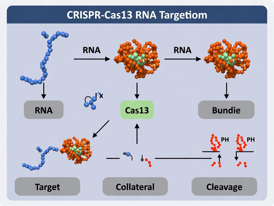
CRISPR-Cas13 systems, particularly Cas13a (C2c2) and Cas13d, have revolutionized programmable RNA targeting for diagnostics, RNA biology, and therapeutic applications. The core thesis of contemporary research posits that while the target-activated, non-specific trans-cleavage (or "collateral") activity of Cas13 is a powerful tool for nucleic acid detection (e.g., SHERLOCK), it represents a significant and complex challenge for in vivo therapeutic applications. This "Collateral Effect" on non-target RNAs introduces potential cytotoxicity, off-target transcript modulation, and unpredictable biological outcomes. This whitepaper provides a technical dissection of the collateral effect, synthesizing current mechanistic understanding, quantitative profiling data, and methodologies for its study and mitigation, framing it within the broader thesis of leveraging versus constraining Cas13's inherent biochemistry.
Mechanism of Cas13 Activation andTrans-Cleavage
Upon formation of a crRNA-guided ternary complex with its cognate target RNA, Cas13 undergoes a conformational activation. This activates its two Higher Eukaryotes and Prokaryotes Nucleotide-binding (HEPN) domains, which form a non-specific RNase active site. The activated state catalyzes the indiscriminate cleavage of nearby single-stranded RNA (ssRNA) molecules, regardless of sequence complementarity. This collateral activity is sustained as long as the activated ternary complex remains intact.
Diagram 1: Cas13 Activation and Collateral Cycle
Quantitative Profiling of Collateral RNA Cleavage
Recent studies have quantified collateral activity in vitro and in cellular models using transcriptomics (RNA-seq). Key metrics include the percentage of transcriptome depletion, sequence/structure biases, and kinetic parameters.
Table 1: Quantification of Cas13d Collateral Activity in Mammalian Cells
| Study (Year) | Cas13 Variant | Delivery | % Transcriptome Reduced (>2-fold) | Notable Bias | Key Finding |
|---|---|---|---|---|---|
| Kushawah et al. (2020) | RfxCas13d (WT) | Plasmid | ~15-25% | Minimal sequence bias | Broad transcriptome-wide knockdown. |
| Mahas et al. (2021) | RfxCas13d (WT) | mRNA | 10-30% | Slight AU-rich preference | Collateral extent correlates with target expression level. |
| Hypothetical Engineered | RfxCas13d (HEPN-) | mRNA | <0.5% | None | Catalytic dead control. |
| Hypothetical Engineered | PspCas13b (WT) | RNP | 5-20% | Structured RNA resistant | Collateral attenuated in dense RNP granules. |
Table 2: In Vitro Kinetic Parameters of Cas13 Trans-Cleavage
| Cas13 Subtype | Target Activation kcat (min-1) | Collateral kcat (min-1) | Reported Processivity | Primary Application |
|---|---|---|---|---|
| LwaCas13a | ~1200 | ~1200 (per complex) | High; bursts of ~1000 cuts | SHERLOCK diagnostics |
| PsmCas13b | ~950 | ~950 (per complex) | Very High | High-sensitivity detection |
| RfxCas13d | ~1800 | ~1800 (per complex) | Moderate-High | In vivo RNA targeting |
Experimental Protocols for Assessing Collateral Cleavage
1In VitroFluorescent Reporter Assay for Collateral Kinetics
Purpose: To quantify the rate and magnitude of Cas13's collateral RNase activity upon target recognition. Reagents: See "The Scientist's Toolkit" below. Procedure:
- Assembly: In a black 384-well plate, mix 50 nM purified Cas13 protein, 50 nM crRNA (target-specific), and 1 nM target RNA in 1X Reaction Buffer. Incubate at 37°C for 10 min.
- Initiation: Add the collateral RNA substrate (100 nM ssRNA reporter with fluorescent dye (FAM) and quencher (BHQ1) at opposite ends) to each well. Final reaction volume: 20 µL.
- Measurement: Immediately transfer plate to a real-time PCR instrument or fluorescent plate reader. Monitor FAM fluorescence (excitation 485 nm, emission 520 nm) every 30 seconds for 1-2 hours at 37°C.
- Analysis: Plot fluorescence vs. time. Calculate the time to reach 50% maximal fluorescence (T50) or initial velocity (RFU/min) as a measure of collateral activity.
Diagram 2: In Vitro Collateral Kinetics Workflow
Cellular RNA-seq for Transcriptome-Wide Off-Target Analysis
Purpose: To identify and quantify non-target RNA degradation in cells expressing active Cas13. Procedure:
- Cell Transfection: Transfect cultured mammalian cells (e.g., HEK293T) with plasmids encoding: a) Wild-type (WT) Cas13 + target-specific crRNA, b) Catalytically dead (d)Cas13 + crRNA, c) Non-targeting crRNA control. Use triplicates.
- Harvest: 48 hours post-transfection, wash cells and lyse in TRIzol. Isolate total RNA following standard protocols.
- Library Prep & Sequencing: Deplete ribosomal RNA. Prepare stranded RNA-seq libraries (e.g., using Illumina TruSeq). Sequence on a Next-Gen platform to a depth of ~30-40 million reads per sample.
- Bioinformatic Analysis:
- Align reads to the reference genome (e.g., STAR aligner).
- Quantify gene expression (e.g., using featureCounts, RSEM).
- Perform differential expression analysis (e.g., DESeq2) comparing WT Cas13 sample vs. dCas13 control.
- Identify significantly downregulated genes (adjusted p-value < 0.05, log2 fold change < -1) as putative collateral cleavage victims. Exclude the intended target gene from this list.
The Scientist's Toolkit: Key Research Reagent Solutions
Table 3: Essential Reagents for Collateral Effect Research
| Reagent / Material | Supplier Examples | Function in Experiment |
|---|---|---|
| Purified Cas13 Proteins (WT & catalytically dead) | IDT, Thermo Fisher, MCLAB, in-house purification | Core enzyme for in vitro kinetics and mechanistic studies. Catalytically dead mutant (dCas13) is the critical negative control. |
| Synthetic crRNAs & Target RNAs | IDT, Dharmacon, Trilink | Define target specificity. High-purity, modified RNAs (e.g., 2'-O-methyl) enhance stability for cellular assays. |
| Fluorescent Quenched RNA Reporters | Biosearch Technologies, IDT | Standardized substrate to measure collateral cleavage rate in vitro (e.g., FAM-dArUdArUdA-BHQ1). |
| RNase Inhibitor (SUPERase•In) | Thermo Fisher | Protects reaction components from non-specific RNase degradation in in vitro assays, ensuring signal specificity. |
| Stranded Total RNA Library Prep Kits | Illumina, NEB, Takara | Enable preparation of sequencing libraries from total RNA to assess transcriptome-wide changes. |
| RiboCop rRNA Depletion Kit | Lexogen | Efficient removal of ribosomal RNA from total RNA samples prior to RNA-seq, enriching for mRNA and non-coding RNA. |
| Lipofectamine MessengerMAX | Thermo Fisher | High-efficiency transfection reagent for delivering Cas13 mRNA and crRNA into mammalian cells for cellular assays. |
| dCas13-APEX2 Proximity Labeling Constructs | Addgene (plasmid deposits) | Tools for identifying the immediate molecular neighborhood and potential substrates of activated Cas13 in cells. |
Mitigation Strategies and Engineered Variants
Current research within the thesis framework focuses on engineering Cas13 to decouple precise targeting from collateral activity.
- HEPN Domain Mutants: Partial or allosteric mutations that reduce trans- while retaining cis-cleavage.
- Conditional Destabilization: Fusing Cas13 to destabilizing domains (DD) controlled by small molecules to limit active complex lifetime.
- Subcellular Localization: Tethering Cas13 to specific organelles (e.g., mitochondria) to restrict its collateral radius.
- High-Fidelity (HiFi) Variants: Directed evolution has yielded Cas13 variants (e.g., Cas13b HiFi) with markedly reduced collateral activity while maintaining on-target potency, representing a promising path for therapeutics.
Diagram 3: Strategies to Mitigate Collateral
The CRISPR-Cas13 system represents a paradigm shift from DNA-targeting CRISPR systems to programmable RNA recognition and cleavage. Within the broader thesis on the CRISPR-Cas13 mechanism of RNA targeting and collateral (trans) cleavage, understanding the diversity within the Cas13 family is crucial. This guide provides a comparative analysis of the three primary subtypes—Cas13a (formerly C2c2), Cas13b, and Cas13d—focusing on their structural features, functional mechanisms, ortholog diversity, and experimental applications in research and therapeutic development.
Core Structural and Functional Diversity
Cas13 proteins are RNA-guided RNA endonucleases. Upon crRNA-guided target RNA recognition, they undergo conformational activation, cleaving the target and promiscuously degrading nearby non-target RNA (collateral cleavage). Key differences among subtypes dictate their application.
Quantitative Comparison of Core Features
Table 1: Comparative Features of Major Cas13 Subtypes
| Feature | Cas13a (C2c2) | Cas13b | Cas13d |
|---|---|---|---|
| Prototypical Orthologs | LbuCas13a, LwaCas13a | PspCas13b, PguCas13b | RfxCas13d (CasRx) |
| Size (aa, approx.) | ~1250-1300 | ~1100-1200 | ~930-1000 |
| crRNA Length (nt) | 64-66 | ~100 | ~70 |
| Direct Repeat (DR) Structure | 5' 28-nt DR, 3' stem-loop | 5' 36-nt DR, 3' stem-loop | 5' 30-nt DR, 3' stem-loop |
| PFS/PAM Requirement | 3' Protospacer Flanking Site (PFS), prefers 'A', 'U', or none (ortholog-dependent) | 5' PFS, 'D' (A/G/U) for some orthologs | No PFS requirement for RfxCas13d |
| Collateral Activity | High | Moderate to High (ortholog-dependent) | Low/Undetectable in mammalian cells |
| Key Domains | 2 HEPN domains, HELICAL1, HELICAL2 | 2 HEPN domains, 1 or 2 HEPN-associated domains | 2 HEPN domains, compact architecture |
| Primary Applications | RNA detection (SHERLOCK), knockdown, viral defense | RNA knockdown, detection, bacterial RNA targeting | Preferred for mammalian RNA knockdown due to high specificity, small size. |
Table 2: Ortholog-Specific Properties and Performance Metrics
| Ortholog | Subtype | PFS | Reported in vitro Collateral Cleavage Rate (k_cat, min⁻¹) | Mammalian Cell Knockdown Efficiency (%) | Key Reference (Example) |
|---|---|---|---|---|---|
| LwaCas13a | Cas13a | 3' non-G | ~1200 | 50-70 | Abudayyeh et al., 2017 |
| LbuCas13a | Cas13a | 3' non-G | ~1000 | 40-60 | Cox et al., 2017 |
| PspCas13b | Cas13b | 5' D (A/G/U) | ~900 | 60-80 | Smargon et al., 2017 |
| RfxCas13d | Cas13d | None | Not detectable in cells | >90 | Konermann et al., 2018 |
| EsCas13d | Cas13d | None | Low in vitro | >85 | Yan et al., 2018 |
Detailed Experimental Protocols
Standardized protocols are essential for comparing Cas13 ortholog function.
Protocol:In VitroCollateral Cleavage Assay (for SHERLOCK-like Detection)
Objective: Quantify collateral RNAse activity upon target RNA recognition. Reagents:
- Purified Cas13 protein (e.g., LbuCas13a, PspCas13b).
- In vitro transcribed crRNA targeting a specific sequence.
- Target RNA (synthetic or transcribed).
- Fluorescent-quenched RNA reporter (e.g., FAM-UU-r(BHQ1)).
- Reaction Buffer: 20 mM HEPES pH 6.8, 50 mM KCl, 5 mM MgCl₂, 5% glycerol, 1 mM DTT.
- Plate reader or real-time PCR machine for fluorescence measurement.
Procedure:
- Complex Formation: Pre-incubate 50 nM Cas13 with 60 nM crRNA in 1x Reaction Buffer for 10 min at 37°C.
- Reaction Setup: In a 96-well plate, mix the complex with 500 nM fluorescent reporter. Initiate the reaction by adding target RNA at varying concentrations (e.g., 0-100 nM).
- Kinetic Measurement: Immediately transfer plate to a fluorimeter. Measure fluorescence (Ex: 485 nm, Em: 535 nm) every 30 seconds for 1-2 hours at 37°C.
- Data Analysis: Calculate the initial velocity (V₀) of fluorescence increase. Plot V₀ vs. target RNA concentration to determine activation kinetics. Compare orthologs under identical conditions.
Protocol: Mammalian Cell RNA Knockdown Efficiency Evaluation
Objective: Compare endogenous mRNA knockdown efficiency of different Cas13 orthologs. Reagents:
- Mammalian expression plasmids for Cas13 orthologs (NLS-tagged).
- U6-driven crRNA expression plasmid targeting a housekeeping gene (e.g., PPIB).
- HEK293T cells.
- Transfection reagent (e.g., PEI, Lipofectamine 3000).
- RT-qPCR reagents (RNA extraction kit, cDNA synthesis kit, SYBR Green master mix).
Procedure:
- Transfection: Seed HEK293T cells in 24-well plates. Co-transfect 500 ng Cas13 plasmid + 250 ng crRNA plasmid per well. Include controls (Cas13 only, crRNA only).
- Harvest: 48-72 hours post-transfection, lyse cells and extract total RNA. Treat with DNase I.
- RT-qPCR: Synthesize cDNA from 500 ng total RNA. Perform qPCR for the target gene and a stable control gene (e.g., GAPDH).
- Analysis: Calculate % knockdown using the 2^(-ΔΔCt) method relative to control (Cas13 only) transfected cells. Perform triplicate biological replicates.
- Specificity Check: Run RNA-seq on high-efficiency candidates (e.g., RfxCas13d) to assess off-target transcriptome effects.
Visualizing Cas13 Mechanism and Experimental Workflows
Diagram 1: Cas13 RNA Targeting and Collateral Cleavage Mechanism
Diagram 2: In Vitro Collateral Cleavage Assay Workflow
The Scientist's Toolkit: Research Reagent Solutions
Table 3: Essential Reagents for Cas13 Research
| Item | Function & Description | Example Supplier/Product (for illustration) |
|---|---|---|
| Recombinant Cas13 Proteins | Purified enzymes for in vitro characterization, detection assay development. | IDT (Alt-R S.p. Cas13a), GenScript (Custom expression/purification). |
| Cas13 Expression Plasmids | Mammalian, bacterial, or AAV-backbone vectors for cellular delivery and knockdown studies. | Addgene (pC0043: RfxCas13d, pC0066: LwaCas13a). |
| crRNA Cloning/Expression Vectors | U6-promoter plasmids for mammalian crRNA expression; T7-promoter templates for in vitro transcription. | Addgene (pC0046: Rfx crRNA vector). |
| Synthetic crRNAs | Chemically synthesized, pre-validated crRNAs for rapid in vitro or ex vivo experiments. | IDT (Alt-R CRISPR-Cas13 crRNA), Synthego. |
| Fluorescent RNA Reporters | Quenched ssRNA probes (FAM/BHQ1) for real-time detection of collateral cleavage activity. | IDT (Alt-R Cas13 Reporter), Biosearch Technologies. |
| Detection Kits (SHERLOCK) | Optimized lyophilized or liquid master mixes for diagnostic nucleic acid detection. | Mammoth Biosciences (DETECTR Kit), Sherlock Biosciences. |
| Nuclease-Free RNA Standards | Quantified, synthetic RNA targets for assay calibration and positive controls. | Twist Bioscience (Synthetic RNA controls). |
| High-Sensitivity RNA Assay Kits | For measuring low-abundance RNA in knockdown efficiency studies (RT-qPCR, RNA-seq). | Bio-Rad (iScript cDNA synthesis), Illumina (RNA Prep with Enrichment). |
| AAV Serotype Vectors | For in vivo delivery of compact Cas13d systems. | PackGene (AAV production services), Vigene Biosciences. |
| Collateral Activity Inhibitors | Small molecules or engineered proteins to modulate collateral effects for therapeutic safety. | Research-grade (e.g., engineered anti-CRISPR AcrVI proteins). |
Cas13a, Cas13b, and Cas13d offer a toolkit with distinct properties. Cas13a remains a cornerstone for sensitive in vitro detection. Cas13b provides robust knockdown in various contexts. Cas13d, with its compact size, high specificity, and minimal collateral activity in cells, is the leading candidate for therapeutic RNA knockdown applications. Future research within the broader thesis will focus on engineering next-generation Cas13 variants with abolished collateral activity, expanded targeting range, and evolved orthologs for in vivo precision medicine, leveraging the comparative framework established here.
Natural Biological Role and Evolutionary Origins of Cas13 Systems
Within the broader thesis on the CRISPR-Cas13 mechanism of RNA targeting and collateral cleavage, understanding the natural biological role and evolutionary origins of these systems is fundamental. Cas13, a Class 2 type VI CRISPR-Cas effector, naturally functions as an adaptive immune system in prokaryotes, providing defense against mobile genetic elements like RNA phages and plasmids. Its evolutionary journey from a putative ancestral nuclease to a specialized RNA-targeting system with unique collateral RNase activity underpins its current utility and limitations in research and therapeutic development.
Natural Biological Role
Cas13 systems operate as RNA-guided, RNA-targeting immune complexes in bacteria and archaea. Upon transcription of the CRISPR array, the mature crRNA guides the Cas13 protein to complementary foreign RNA sequences. Target recognition activates the two Higher Eukaryotes and Prokaryotes Nucleotide-binding (HEPN) domains within Cas13, triggering precise cleavage of the target and promiscuous, non-specific degradation of nearby bystander RNA (collateral cleavage). This collateral activity can induce cell dormancy or death, functioning as an abortive infection mechanism to limit the spread of the viral infection within a bacterial population.
Table 1: Key Functional Characteristics of Canonical Cas13 Subtypes
| Subtype | Primary Effector | Size (aa) | Required Protospacer Flanking Sequence (PFS) | Natural Putative Function |
|---|---|---|---|---|
| Type VI-A | Cas13a (C2c2) | ~1250 | 5' non-G (for some orthologs) | Defense against RNA phages; abortive infection. |
| Type VI-B | Cas13b | ~1150 | 5' and 3' PFS present | Defense against RNA phages; plasmid interference. |
| Type VI-C | Cas13c | ~1050 | None reported | Defense against RNA phages. |
| Type VI-D | Cas13d | ~930 | None | Defense against RNA phages; compact genetic locus. |
Evolutionary Origins and Classification
Cas13 proteins are classified within Class 2 (single multi-domain effector) Type VI CRISPR-Cas systems. Phylogenetic analyses suggest Cas13 evolved from a family of ancestral HEPN-domain containing RNases, unrelated to the RuvC/HNH endonuclease fold of Cas9. The core HEPN domains, responsible for RNase activity, are conserved across all Cas13 variants and related to widespread RNases found in toxin-antitoxin and other defense systems. This indicates an evolutionary path where a standalone non-specific RNase acquired CRISPR-associated guide RNA recognition modules (Helical-1 and Helical-2 domains) to become a programmable, specific immune effector. The diversity of subtypes (VI-A to VI-D) reflects adaptation to different genomic contexts and selective pressures.
Table 2: Evolutionary and Genomic Features of Cas13 Systems
| Feature | Evolutionary Implication | Supporting Evidence |
|---|---|---|
| HEPN Domain Conservation | Descended from ancient, ubiquitous RNase fold. | Structural alignment with TnpB RNases and prokaryotic toxins. |
| Locus Architecture | Frequent association with WYL-domain accessory proteins (e.g., WYL1 in Cas13b). | Suggests regulatory mechanisms evolved to control collateral activity. |
| Size Variation (930-1300 aa) | Diversification and miniaturization for efficiency. | Cas13d is the smallest, likely a highly derived, compact form. |
| PFS Diversity | Adaptation to recognize specific viral sequence patterns. | PFS requirements vary between subtypes and orthologs. |
Core Experimental Protocols for Functional Analysis
Protocol: Assessing Natural Antiviral DefenseIn Vivo
Objective: To demonstrate Cas13's role in defending against RNA phage infection in its native bacterial host. Methodology:
- Strain Construction: Clone the endogenous Cas13 CRISPR locus (including effector gene, CRISPR array, and accessory genes) into a plasmid vector. Transform into a naive bacterial strain lacking a native CRISPR system. Include a control strain with a catalytically inactive Cas13 (HEPN mutations, e.g., RxxxxH).
- Phage Challenge: Grow Cas13-expressing and control strains to mid-log phase. Infect with a series of dilutions of a known RNA phage (e.g., MS2). Co-incubate for a set period (e.g., 4 hours).
- Plaque Assay: Plate the infected culture on a lawn of a susceptible indicator bacterial strain. Incubate overnight.
- Data Analysis: Count plaques. Calculate plaque-forming units (PFU/mL). Defense is quantified as the reduction in PFU/mL in the active Cas13 strain versus the inactive control.
Protocol: Quantifying Collateral RNase ActivityIn Vitro
Objective: To measure the kinetics and specificity of target-activated non-specific RNA cleavage. Methodology:
- Reaction Setup: Purify recombinant Cas13 protein and transcribe its cognate crRNA. Assemble the ribonucleoprotein (RNP) complex.
- Substrate Preparation: Synthesize a fluorescently quenched (FAM/BHQ) target RNA oligo perfectly matching the crRNA spacer. For bystander RNA, use a synthetic, non-complementary RNA oligo labeled with a distinct fluorophore (e.g., Cy5) or total bacterial RNA stained with an intercalating dye (e.g., SYBR Green II).
- Kinetic Measurement: Use a real-time fluorescence plate reader. Mix RNP with target and bystander substrates simultaneously. Monitor fluorescence increase (dequenching) in both the target (FAM, 518 nm) and bystander (Cy5, 670 nm; or SYBR, 525 nm) channels every 30 seconds for 1-2 hours.
- Data Analysis: Plot fluorescence vs. time. Calculate initial reaction velocities (V0) and time to half-maximal cleavage for both target and collateral substrates.
Diagrams
Diagram 1: Cas13 antiviral function with collateral effect.
Diagram 2: Proposed evolutionary origin of Cas13 systems.
The Scientist's Toolkit: Key Research Reagent Solutions
Table 3: Essential Reagents for Cas13 Biology Research
| Reagent | Function/Description | Example Product/Catalog |
|---|---|---|
| Recombinant Cas13 Protein (Purified) | Catalytic effector for in vitro cleavage assays. Requires HEPN domain integrity. | LbuCas13a, PspCas13b, RfxCas13d (NEB, IDT, custom expression) |
| crRNA Synthesis Kit | For generating guide RNA complementary to target sequence. Requires 5' handle for Cas13 loading. | Custom synthetic crRNA (IDT, Sigma), T7 *in vitro transcription kit (NEB)* |
| Fluorescent RNA Oligonucleotides | Labeled target and bystander substrates for real-time kinetic measurement of cleavage. | FAM/BHQ-labeled target RNA, Cy5-labeled bystander RNA (IDT, Horizon) |
| HEPN-Domain Mutant Cas13 (dCas13) | Catalytically dead control (e.g., R->A/H->A mutations). Used to distinguish guide-binding from cleavage effects. | Plasmids available from Addgene (e.g., #109049 for LwaCas13a) |
| RNA Purification Kit (RNase-free) | Critical for isolating high-quality RNA for collateral activity assays and NGS analysis. | QIAGEN RNeasy, Zymo RNA Clean & Concentrator |
| In Vivo Phage Stock | RNA bacteriophage for challenge experiments in native or heterologous hosts. | MS2, Qβ, PP7 (ATCC, research labs) |
| WYL-domain Accessory Proteins | For studying regulation of Cas13b/d systems in vitro and in vivo. | Recombinant WYL1, Csx27/28 proteins (custom expression) |
Harnessing the Power: Step-by-Step Guide to Cas13 Applications in Research and Diagnostics
Within the broader study of the CRISPR-Cas13 mechanism of RNA targeting and its non-specific collateral cleavage activity, experimental design is paramount. This guide provides a technical framework for selecting Cas13 orthologs and designing crRNAs, critical for applications in RNA detection, knockdown, and therapeutic development.
Key Cas13 Orthologs: Characteristics and Selection Criteria
Cas13 orthologs vary in size, crRNA requirements, collateral activity, and temperature sensitivity, influencing their suitability for specific applications. The following table summarizes quantitative characteristics of the most commonly used orthologs.
Table 1: Comparative Analysis of Common Cas13 Orthologs
| Ortholog | Size (aa) | Pre-crRNA Length | Direct Repeat Sequence | PFS Preference | Reported Collateral Activity | Optimal Temp. | Primary Applications |
|---|---|---|---|---|---|---|---|
| Cas13a (LshCas13a) | ~1250 | 64 nt | 5'-GUUCACUGCCGUAUAGGCAGCUAGAAU-3' | 3' of target: A, U (non-A for Lwa) | High | 37°C | SHERLOCK, RNA knockdown |
| Cas13b (PspCas13b) | ~1150 | 64 nt | 5'-AAAUUCUCUCUAGUAGCAUGUAAAAAC-3' | 3' of target: D (A,G,U) | Moderate/High | 37°C | RNA editing (RESCUE), knockdown |
| Cas13d (RfxCas13d) | ~967 | 63 nt | 5'-AAACACCCTACCAATGGACAGCTTCGG-3' | None | Moderate | 37°C | In vivo RNA knockdown, therapeutics |
| Cas13a (LwaCas13a) | ~1250 | 64 nt | 5'-GUUCACUGCCGUAUAGGCAGCUAGAAU-3' | 3' of target: Non-A | High | 37°C | SHERLOCK (high sensitivity) |
| Cas13a (CcaCas13b) | ~1120 | 64 nt | 5'-UAAAUUUCAACCCGUGUGUGGUGGGACU-3' | 5' of target: H (A,C,U) | High | 37-50°C | Thermostable diagnostics |
crRNA Design Rules and Best Practices
Effective crRNA design is critical for on-target efficiency and minimization of off-target effects.
3.1 Core Design Parameters:
- Spacer Length: Typically 28-30 nucleotides for most orthologs. Shorter spacers (20-24 nt) can increase specificity but may reduce activity.
- Spacer Sequence: Must be complementary to the target RNA. Avoid extensive secondary structure within the spacer.
- Direct Repeat (DR): The ortholog-specific handle must be correctly appended to the 5' (Cas13a/d) or 3' (Cas13b) end of the spacer.
- Protospacer Flanking Site (PFS): Respect the ortholog-specific nucleotide preference (see Table 1). Cas13d has no known PFS, offering greater target flexibility.
3.2 Rules for Minimizing Off-Targeting:
- Mismatches in the spacer seed region (typically nucleotides 3-10 from the 3' end of the spacer) severely reduce cleavage.
- Use predictive algorithms (e.g., Cas13design, FlashFold) to assess crRNA folding and target accessibility.
- Empirically validate multiple crRNAs per target.
Table 2: crRNA Design Specifications by Ortholog
| Ortholog | Spacer Length (nt) | DR Position | Key Design Consideration |
|---|---|---|---|
| Cas13a | 28 | 5' | Ensure correct PFS (A/U) 3' of target. |
| Cas13b | 30 | 3' | Ensure correct PFS (D) 3' of target. |
| Cas13d | 22-30 | 5' | No PFS constraint. 22-nt spacers often used for in vivo efficiency. |
Key Experimental Protocols
Protocol 1: In Vitro Collateral Cleavage Assay (for Diagnostic Development) Purpose: To quantify the collateral RNase activity of a Cas13 ortholog upon target RNA recognition.
- Purified Component Assembly: Combine in a reaction: 50 nM purified Cas13 ortholog, 50 nM in vitro-transcribed crRNA, 5 nM target RNA transcript, and 500 nM quenched fluorescent RNA reporter (e.g., FAM-UUUU-BHQ1).
- Buffer Conditions: Use reaction buffer (20 mM HEPES pH 6.8, 50 mM KCl, 5 mM MgCl2, 1 mM DTT, 5% glycerol).
- Kinetic Measurement: Load mixture into a real-time PCR instrument or fluorometer. Measure fluorescence (Ex/Em: 485/535 nm for FAM) every 1-2 minutes for 1-2 hours at 37°C.
- Analysis: Calculate initial reaction rates (RFU/min) to compare ortholog collateral activity or crRNA efficiency.
Protocol 2: Cellular RNA Knockdown Validation Purpose: To evaluate Cas13-mediated RNA targeting in mammalian cells.
- Delivery: Co-transfect HEK293T cells (or relevant cell line) with two plasmids: a) expressing the Cas13 ortholog, and b) expressing the crRNA from a U6 promoter. Include a non-targeting crRNA control.
- Target Co-transfection: Include a plasmid expressing a tagged target RNA if targeting endogenous transcripts is not feasible.
- Harvest: 48-72 hours post-transfection, lyse cells.
- Validation: Quantify target RNA levels via RT-qPCR. Assess protein-level knockdown via western blot if applicable.
- Specificity Check: Perform RNA-Seq or probe for related transcripts to assess off-target collateral effects in cells.
Visualizing the Cas13 Targeting and Collateral Pathway
Diagram 1: Cas13 Activation and Dual Cleavage Pathways (76 chars)
The Scientist's Toolkit: Key Research Reagent Solutions
Table 3: Essential Reagents for Cas13 Research
| Reagent/Catalog | Function & Explanation |
|---|---|
| Purified Recombinant Cas13 Protein (e.g., LwaCas13a) | Core enzyme for in vitro assays (collateral cleavage, diagnostics). Purity is critical for high signal-to-noise. |
| T7 RNA Polymerase Kit | For high-yield in vitro transcription of target RNA and crRNA templates. |
| Fluorescent Quenched RNA Reporter (FAM-UUUU-BHQ1) | Universal substrate for detecting Cas13 collateral activity; cleavage unquenches fluorophore. |
| RNase Inhibitor (Murine or Human) | Essential for preventing non-specific RNA degradation in all pre-assembly reaction steps. |
| CrRNA Cloning Vector (e.g., pC013 or pC004) | Plasmid backbone for expressing crRNA from a U6 promoter in mammalian cells. |
| Cas13 Mammalian Expression Vector | Plasmid for constitutive or inducible expression of codon-optimized Cas13 orthologs (e.g., pC009 for LwaCas13a). |
| Nucleofection or Lipofection Kit | For efficient delivery of RNP complexes or plasmids into hard-to-transfect cell lines. |
| RT-qPCR Kit with RNA-specific Probes | Gold-standard for validating target RNA knockdown in cellular experiments. |
This technical guide details the protocols for three leading CRISPR-based diagnostic platforms: SHERLOCK, DETECTR, and CARMEN. These methods are built upon the foundational research into the CRISPR-Cas13 mechanism of RNA targeting and collateral cleavage. Cas13, upon activation by its target RNA sequence, exhibits promiscuous RNase activity, cleaving surrounding non-target reporter RNA molecules. This "collateral cleavage" forms the core signal amplification principle for SHERLOCK and CARMEN. Similarly, Cas12a's collateral ssDNA cleavage is leveraged by DETECTR. This whitepaper frames these diagnostics within the ongoing thesis research exploring the kinetics, specificity, and optimization of this collateral effect for ultrasensitive pathogen detection and genotyping.
Table 1: Comparative Overview of CRISPR Diagnostic Platforms
| Feature | SHERLOCK (v2) | DETECTR | CARMEN |
|---|---|---|---|
| CRISPR Enzyme | Cas13a (LwaCas13a, RfxCas13d) | Cas12a (LbCas12a, AsCas12a) | Cas13 (Various, e.g., LwaCas13a) |
| Target Nucleic Acid | RNA | DNA (ss/ds) | RNA & DNA (Multiplexed) |
| Pre-amplification | RPA or RT-RPA | RPA | PCR or RPA |
| Core Detection Mechanism | Collateral cleavage of fluorescent RNA reporter | Collateral cleavage of fluorescent ssDNA reporter | Collateral cleavage of fluorescent RNA reporter in microwell |
| Readout Modality | Fluorescence (Lateral flow strip or plate reader) | Fluorescence (Lateral flow strip or plate reader) | Fluorescence via microfluidic array imaging |
| Reported Sensitivity | ~2 aM (attomolar) | ~aM to single-digit fM | High-plex detection at aM-fM |
| Key Multiplexing Capacity | ~4 targets (using orthogonal Cas13 proteins & reporters) | ~2-3 targets | >4,500 targets in a single array (theoretical) |
| Time to Result | ~60-90 minutes | ~45-90 minutes | ~5-8 hours (including array fabrication) |
Table 2: Quantitative Performance Metrics from Recent Studies
| Platform (Target) | Limit of Detection (LoD) | Clinical Sensitivity/Specificity | Key Citation (Example) |
|---|---|---|---|
| SHERLOCK (SARS-CoV-2 RNA) | 42 copies/µL | 100% / 100% (in selected cohort) | Joung et al., NEJM, 2020 |
| DETECTR (HPV16 in cell lines) | ~0.75 copies/µL | 95% / 100% (vs. sequencing) | Chen et al., Science, 2018 |
| CARMEN (169 human-associated viruses) | 1-10 copies/µL per pathogen | Enables comprehensive viral panel detection | Ackerman et al., Nature, 2020 |
Detailed Experimental Protocols
SHERLOCK Protocol for Viral RNA Detection
Principle: Target RNA is isothermally amplified via Recombinase Polymerase Amplification (RPA) with a T7 promoter sequence incorporated. The amplicon is then transcribed by T7 RNA polymerase, generating the target RNA that activates Cas13. Activated Cas13 cleaves a fluorescently quenched RNA reporter, generating a detectable signal.
Key Reagents & Materials:
- Sample: Purified RNA or crude lysate.
- Amplification: RT-RPA pellets (TwistAmp Basic kit).
- Enzymes: T7 RNA polymerase, RNase Inhibitor, LwaCas13a or RfxCas13d.
- Reporter: Fluorescent Quenched RNA Reporter (e.g., FAM-UUUU-BHQ1).
- Buffer: Optimized Cas13 reaction buffer (e.g., 20 mM HEPES, 60 mM NaCl, 6 mM MgCl2, pH 6.8).
Procedure:
- Isothermal Amplification (35 min, 42°C):
- Prepare a 50 µL RT-RPA reaction containing: 5 µL sample RNA, 29.5 µL rehydration buffer, 2.5 µL forward primer (10 µM), 2.5 µL reverse primer with T7 promoter (10 µM), and 10 µL magnesium acetate.
- Incubate at 42°C for 35 minutes.
- Cas13 Detection Reaction (30-60 min, 37°C):
- Prepare a 20 µL detection mix containing: 1 µL Cas13 enzyme (100 nM), 1 µL reporter RNA (1 µM), 1 µL T7 RNA polymerase, 0.5 µL RNase Inhibitor, and 16.5 µL reaction buffer.
- Transfer 2 µL of the RPA amplicon into the detection mix.
- Incubate at 37°C and monitor fluorescence (FAM channel: Ex/Em ~485/535 nm) in a real-time PCR machine or plate reader at 1-2 minute intervals.
- Lateral Flow Readout (Alternative):
- After detection reaction, apply mixture to a lateral flow strip designed to capture biotin-labeled and FAM-labeled cleavage products.
DETECTR Protocol for DNA Target Detection
Principle: Target DNA is amplified isothermally via RPA. The dsDNA amplicon activates Cas12a, which then exhibits collateral cleavage activity against a fluorescently quenched ssDNA reporter.
Key Reagents & Materials:
- Sample: Purified DNA.
- Amplification: RPA pellets (TwistAmp Basic kit).
- Enzymes: LbCas12a.
- Reporter: Fluorescent Quenched ssDNA Reporter (e.g., FAM-TTATT-BHQ1).
- Buffer: NEBuffer 2.1 or similar.
Procedure:
- Isothermal Amplification (30 min, 37°C):
- Prepare a 50 µL RPA reaction as per manufacturer's instructions with sample DNA and target-specific primers.
- Incubate at 37°C for 30 minutes.
- Cas12a Detection Reaction (15-30 min, 37°C):
- Prepare a 20 µL detection mix containing: 1 µL LbCas12a (100 nM), 1 µL crRNA (100 nM), 1 µL ssDNA reporter (1 µM), and 17 µL reaction buffer.
- Transfer 2 µL of the RPA amplicon into the detection mix.
- Incubate at 37°C and monitor fluorescence in real-time. Signal generation is typically faster than Cas13.
CARMEN Protocol for Multiplexed Detection
Principle: Sample nucleic acids are amplified and transcribed (if needed). Each sample is then mixed with a unique color code (fluorescent dye) and loaded into a microwell of a microfluidic array. Each assay (Cas13/crRNA/reporter mix) is loaded with a different color code into another set of wells. The system pairs samples and assays via droplet pairing and coalescence, enabling thousands of simultaneous Cas13 detection reactions in nanoliter droplets.
Key Reagents & Materials:
- Sample Prep: Primers with T7 promoters for PCR/RPA and in vitro transcription.
- Chip Fabrication: Photopolymerizable oil, microfluidic array chips.
- Encoding Dyes: A set of 4-8 spectrally distinct fluorophores (e.g., Alexa Fluor 488, 555, 594, 647) for sample and assay labeling.
- Detection Mix: LwaCas13a, target-specific crRNA, fluorescent RNA reporter.
Procedure:
- Sample and Assay Preparation (2-3 hours):
- Amplify and transcribe all samples to generate target RNA.
- Encode each sample with a unique combination of two fluorescent dyes.
- Prepare assay mixes for each target: Cas13, crRNA, reporter. Encode each assay mix with its own unique two-dye combination.
- Microfluidic Array Loading and Pairing (1-2 hours):
- Use the CARMEN (Combinatorial Arrayed Reactions for Multiplexed Evaluation of Nucleic acids) platform to dispense encoded samples and assays into separate microwells on a microfluidic chip.
- Apply an electric field to pair sample and assay droplets, which then merge.
- Incubation and Imaging (1-2 hours, 37°C):
- Incubate the chip at 37°C to allow Cas13 reactions to proceed.
- Image the entire array using a fluorescence microscope with multiple channels to decode the sample/assay identity and read the positive (reporter cleavage) signal.
Visualization: Experimental Workflows and Mechanisms
Diagram 1: SHERLOCK Workflow from Sample to Signal
Diagram 2: CARMEN Multiplexing Workflow and Encoding Logic
The Scientist's Toolkit: Key Research Reagent Solutions
Table 3: Essential Reagents for CRISPR Diagnostics Development
| Reagent Category | Specific Example(s) | Function in Protocol | Key Consideration for Research |
|---|---|---|---|
| CRISPR Enzymes | Purified LwaCas13a, RfxCas13d (Cas13); LbCas12a, AsCas12a (Cas12) | Core detection protein; collateral nuclease activity. | Orthogonal proteins enable multiplexing. Purity affects background signal. |
| Synthetic crRNAs | Chemically synthesized, target-specific crRNAs with direct repeat. | Guides Cas enzyme to target sequence; defines specificity. | Requires optimization of spacer length (28 nt for Cas13) and minimal off-target homology. |
| Fluorescent Reporters | RNA: FAM-rUrUrUrU-3BHQ; ssDNA: FAM-TTATT-3BHQ | Collateral cleavage substrate; fluorescence de-quenched upon cutting. | Quencher type (BHQ1, BHQ2) and linker length impact signal-to-noise ratio. |
| Isothermal Amplification Kits | TwistAmp Basic RPA/RT-RPA kits (TwistDx/Twist Bioscience) | Rapid, low-temperature amplification of target. | Primer design is critical; must avoid primer-dimers that consume reagents. |
| T7 RNA Polymerase | High-concentration, RNase-free T7 RNA Pol (e.g., NEB) | Transcribes RPA amplicon to RNA for Cas13 detection (SHERLOCK). | High yield is necessary for sensitivity. |
| RNase Inhibitor | Recombinant RNase Inhibitor (e.g., Murine) | Protects RNA reporter and target RNA from degradation. | Essential for robust Cas13 assay performance. |
| Lateral Flow Strips | Milenia HybriDetect strips (for FAM/Biotin detection) | Provides visual, instrument-free readout. | Compatibility with cleavage products must be validated. |
| Microfluidic Encoder Dyes | Alexa Fluor 488, 555, 594, 647 carboxylic acid succinimidyl esters | Uniquely labels sample and assay droplets for CARMEN. | Dyes must be stable, bright, and spectrally distinct. |
| Nuclease-free Buffers | Custom buffers (HEPES, MgCl2, DTT, etc.) | Optimizes enzyme kinetics and stability. | Mg2+ concentration is a critical variable for Cas13 activity. |
Within the broader study of CRISPR-Cas13 mechanisms and collateral cleavage, the Cas13d system has emerged as a powerful, precise tool for RNA targeting without DNA alteration. This whitepaper provides a technical guide to leveraging Cas13d for transcriptional silencing (knockdown) and RNA base editing (via RESCUE and REPAIR systems), focusing on experimental design and implementation for therapeutic development.
Core Mechanisms of Cas13d
Cas13d (e.g., from Ruminococcus flavefaciens XPD3002, known as CasRx) is a Type VI-D CRISPR RNA-guided ribonuclease. Upon binding to its target single-stranded RNA via a crRNA spacer, its two HEPN (Higher Eukaryotes and Prokaryotes Nucleotide-binding) domains are activated for RNA cleavage. A critical area of research is understanding and mitigating its non-specific "collateral" RNase activity upon target recognition, which, while a concern for diagnostics, is typically minimized in eukaryotic cells due to compartmentalization and can be engineered out (e.g., catalytically dead dCas13d) for editing applications.
Application 1: Transcriptional Silencing with Catalytically Active Cas13d
The wild-type Cas13d protein can be programmed to cleave specific mRNA transcripts in the cytoplasm, leading to degradation and effective gene knockdown.
Key Protocol: Cas13d-mediated mRNA Knockdown in Mammalian Cells
Objective: To achieve targeted degradation of a specific mRNA transcript in cultured mammalian cells.
Materials & Reagents:
- Cas13d Expression Plasmid: Mammalian codon-optimized Cas13d (e.g., CasRx) under a strong promoter (EF1α, CAG).
- crRNA Expression Cassette: Typically delivered as a single guide RNA (sgRNA) expressed from a U6 promoter. The spacer sequence (22-28 nt) is designed to target the desired mRNA region, preferably in an accessible, non-structured area like the 3' UTR or coding sequence.
- Delivery Method: Lipofectamine 3000 for HEK293T cells, or appropriate viral vectors (AAV, lentivirus) for primary cells/in vivo.
- Control Plasmids: Non-targeting crRNA and fluorescent reporter (e.g., GFP) for normalization.
Procedure:
- Design & Cloning: Design crRNA spacer sequences complementary to the target mRNA. Clone the spacer into a mammalian sgRNA expression plasmid downstream of a U6 promoter.
- Cell Transfection: Co-transfect the Cas13d expression plasmid and the crRNA plasmid into cells (e.g., 500 ng each per well in a 24-well plate) using a lipid-based transfection reagent.
- Incubation: Harvest cells 48-72 hours post-transfection.
- Validation:
- qRT-PCR: Isolate total RNA, perform reverse transcription, and run qPCR with primers flanking the target site to quantify mRNA knockdown.
- Western Blot: Assess protein level reduction 72-96 hours post-transfection.
Expected Outcome: Effective knockdown (70-95%) at the mRNA level, with corresponding protein reduction.
Application 2: RNA Base Editing with Engineered dCas13d
Catalytically inactive dCas13d serves as a programmable RNA-binding platform. Fused to deaminase domains, it enables precise RNA base editing without permanent genomic changes.
The REPAIR and RESCUE Systems
- REPAIR (RNA Editing for Programmable A to I Replacement): dCas13b fused to the adenosine deaminase domain of ADAR2 (TadA variants) converts adenosine (A) to inosine (I), read as guanosine (G) by cellular machinery.
- RESCUE (RNA Editing for Specific C to U Exchange): An evolved version of the REPAIR deaminase (ADAR2dd E488Q) with cytidine deaminase activity, enabling C-to-U editing.
Recent advances have adapted these editors to the compact Cas13d backbone (dCas13d-ADAR fusions) for improved delivery and efficiency.
Key Protocol: dCas13d-ADAR Fusion for C-to-U RNA Editing (RESCUE)
Objective: To perform specific C-to-U RNA editing on a endogenous transcript in cells.
Materials & Reagents:
- RESCUE Editor Plasmid: Mammalian expression vector encoding dCas13d (HEPN catalytic sites mutated, e.g., RXXXH to AXXXA) fused to the evolved ADAR2 deaminase domain (E488Q).
- Targeting sgRNA Plasmid: U6-driven sgRNA with spacer designed to place the target cytidine within the deaminase window (typically spacer positions 4-8, 3' of the protospacer).
- RNA Extraction & cDNA Synthesis Kit
- Sanger Sequencing or Next-Generation Sequencing (NGS) Supplies for analysis.
Procedure:
- System Design: Design 3-5 sgRNAs targeting the region of interest, predicting the editing window relative to the sgRNA binding site.
- Transfection: Co-transfect the RESCUE editor plasmid and sgRNA plasmid into cells.
- RNA Harvest: Extract total RNA 48 hours post-transfection. Treat with DNase I to remove plasmid DNA contamination.
- Analysis:
- RT-PCR & Sanger Sequencing: Perform RT-PCR on the target region, purify the amplicon, and submit for Sanger sequencing. Analyze chromatograms for C-to-T (U) peak overlaps.
- Deep Sequencing (Gold Standard): Design amplicons for the target site and perform high-throughput sequencing. Use analysis pipelines (e.g., CRISPResso2) to quantify precise editing efficiency and identify potential off-target edits.
Expected Outcome: Site-specific C-to-U conversion with variable efficiency (typically 10-50%, depending on context). Off-target RNA editing should be assessed via transcriptome-wide analysis (e.g., RNA-seq).
Table 1: Comparison of Cas13d RNA-Targeting Systems
| System | Core Enzyme | Primary Activity | Editing Window (relative to spacer 5' end) | Typical Efficiency in Mammalian Cells | Key Outcome |
|---|---|---|---|---|---|
| Cas13d (Knockdown) | Wild-type Cas13d | RNA cleavage (knockdown) | Cleavage site within target region | 70-95% mRNA reduction | Transcript degradation |
| REPAIR (dCas13d-ADAR) | dCas13d + ADAR2dd | A-to-I (G) editing | ~Position 4-8 (3' of protospacer) | 20-80% (highly site-dependent) | A->G point mutation at RNA level |
| RESCUE (dCas13d-ADAR2dd E488Q) | dCas13d + evolved ADAR2dd (E488Q) | C-to-U (T) editing | ~Position 4-8 (3' of protospacer) | 10-50% (highly site-dependent) | C->U point mutation at RNA level |
Table 2: Common Research Reagent Solutions
| Reagent / Material | Example Product / Specification | Primary Function in Experiment |
|---|---|---|
| Cas13d/dCas13d Expression Plasmid | Addgene #109049 (pXR001: CasRx), or custom dCas13d-ADAR fusions | Delivers the effector protein backbone for RNA binding and catalytic/deaminase activity. |
| sgRNA Cloning Backbone | Addgene #109053 (pXR003: U6-sgRNA scaffold) | Allows for efficient insertion and expression of target-specific spacer sequences. |
| Delivery Reagent (in vitro) | Lipofectamine 3000 (Thermo Fisher) | Transient transfection of plasmid DNA into mammalian cell lines. |
| RNA Extraction Kit | TRIzol Reagent or column-based kits (e.g., Qiagen RNeasy) | Isolates high-quality total RNA for downstream qRT-PCR or sequencing analysis. |
| RT-qPCR Master Mix | iTaq Universal SYBR Green One-Step Kit (Bio-Rad) | Quantifies mRNA levels from extracted RNA to assess knockdown efficiency. |
| NGS Library Prep Kit | Illumina DNA Prep or NEBNext Ultra II RNA Library Prep | Prepares amplicons or RNA-seq libraries for deep sequencing to quantify editing and off-targets. |
| Control crRNA/sgRNA | Non-targeting spacer (e.g., targeting GFP or scrambled sequence) | Essential negative control to distinguish specific effects from non-specific cellular responses. |
Visualizations
Title: Cas13d Experimental Workflow Selection
Title: dCas13d-ADAR Fusion Mechanism for RNA Editing
Live-Cell RNA Imaging and Tracking with Catalytically Inactive dCas13
The CRISPR-Cas13 system, unlike DNA-targeting Cas9, is a Class 2, Type VI RNA-guided RNase. Its canonical mechanism involves two core functions: sequence-specific binding and cleavage of target single-stranded RNA (ssRNA) via its HEPN (Higher Eukaryotes and Prokaryotes Nucleotide-binding) domains, and promiscuous collateral trans-cleavage of nearby non-target RNAs upon target recognition. The latter activity, while useful for diagnostic applications, is cytotoxic and unsuitable for live-cell applications. This whitepaper is framed within a broader thesis investigating the precise molecular determinants of Cas13's RNA targeting fidelity and the structural basis of collateral cleavage activation. The development of a catalytically inactive "dead" Cas13 (dCas13), where point mutations (e.g., HEPN domain RxxxxH to AxxxxA) abolish both cis and trans nuclease activities while preserving RNA binding, provides a foundational tool. This enables the repurposing of dCas13 for non-destructive, programmable RNA visualization and tracking, offering unprecedented insights into RNA metabolism, localization, and regulation in living cells—a critical advancement for both basic research and drug development targeting RNA processes.
Core Principles of dCas13-Based Imaging
dCas13 functions as a programmable RNA-binding protein. When fused to a fluorescent protein (e.g., GFP, mCherry), it can be guided by a specific crRNA to label endogenous RNA transcripts. Key advantages over older methods (e.g., MS2-MCP) include direct programmability without genetic engineering of the target RNA and the potential for multiplexing with orthologous Cas13 variants (e.g., PspCas13b, RfxCas13d). The signal intensity is a function of dCas13-crRNA complex abundance, target RNA copy number, and binding turnover kinetics. A critical consideration is the necessity of robust nuclear export signals (NES) on dCas13 fusions for cytoplasmic RNA targeting, and the optimization of crRNA design to ensure high specificity and avoid target site occlusion by RNA-binding proteins.
Table 1: Comparison of Common dCas13 Orthologs for Live-Cell Imaging
| Ortholog | Size (aa) | Preferred PFS* | Guide Length | Key Attributes for Imaging | Typical Fusion Size (kDa) |
|---|---|---|---|---|---|
| dPspCas13b | 1127 | None | 30 nt | High specificity, compact for delivery | ~140 (with GFP) |
| dRfxCas13d | 967 | None | 22-30 nt | Smallest, high efficiency in mammalian cells | ~120 (with GFP) |
| dLwaCas13a | 1228 | 3' H, U | 28 nt | Requires PFS, used in early proof-of-concept | ~150 (with GFP) |
*Protospacer Flanking Sequence requirement.
Table 2: Performance Metrics of dCas13 Imaging Systems (Representative Data)
| Metric | Typical Range/Value | Experimental Condition Notes |
|---|---|---|
| Signal-to-Background Ratio | 5:1 to 30:1 | Dependent on target abundance and crRNA design. |
| Time to Detectable Signal | 1-6 hours post-transfection | Varies with delivery method and expression kinetics. |
| Photostability | Limited by FP half-life; can be improved with tags like HaloTag/SNAP-tag. | Enables longer time-lapse tracking. |
| Reported Spatial Resolution | ~200-300 nm (diffraction-limited) | Can be combined with super-resolution techniques. |
| Multiplexing Capacity (Proof-of-Concept) | Up to 3 colors | Using orthogonal dCas13 proteins with distinct crRNAs and FP colors. |
Detailed Experimental Protocols
Protocol 4.1: Construct Design and Preparation for dCas13 Imaging
- dCas13 Backbone: Start with a mammalian expression plasmid (e.g., pcDNA3.1, pCAG) containing a catalytically inactive Cas13 (e.g., dPspCas13b with mutations R1188A/H1191A).
- Fluorescent Fusion: Clone in-frame, via Gibson Assembly or similar, a fluorescent protein (e.g., EGFP, mNeonGreen) at the Cas13 C-terminus. Include a flexible linker (e.g., GGSGGS x3).
- Localization Signals: For cytoplasmic RNA targets, incorporate a strong NES (e.g., from PKI) between dCas13 and the FP. For nuclear-retained RNA, use an NLS.
- crRNA Expression: Use a U6 promoter-driven plasmid to express the crRNA. The direct repeat sequence is ortholog-specific. Clone the 22-30nt spacer sequence complementary to the target RNA immediately downstream.
Protocol 4.2: Live-Cell Imaging of β-actin mRNA with dPspCas13b-EGFP
Day 1: Cell Seeding
- Seed HeLa or HEK293T cells in a glass-bottom 35-mm imaging dish at 50-70% confluence in complete medium.
Day 2: Transfection
- Prepare two transfection mixes:
- Mix A (1.5µg DNA): 1.0 µg dPspCas13b-EGFP-NES plasmid + 0.5 µg U6-crRNA[β-actin] plasmid.
- Mix B (Control): 1.0 µg dPspCas13b-EGFP-NES plasmid + 0.5 µg U6-crRNA[Scramble] plasmid.
- Use a lipofection reagent (e.g., Lipofectamine 3000) according to manufacturer instructions.
- Incubate cells with complexes for 4-6 hours, then replace with fresh medium.
Day 3-4: Imaging (24-48h post-transfection)
- Transfer dish to a pre-warmed (37°C), CO₂-controlled confocal microscope stage.
- Use a 60x or 100x oil-immersion objective.
- EGFP Imaging: Excite at 488 nm, collect emission at 500-550 nm.
- Acquire Z-stacks (0.5 µm steps) or single optical sections.
- For time-lapse, acquire images every 15-30 seconds for up to 30 minutes to track RNA particle movement. Minimize laser power to reduce photobleaching.
Day 4: Validation (Post-imaging)
- Perform single-molecule RNA FISH against the target RNA on the same cell line to confirm localization patterns observed with dCas13.
Protocol 4.3: Specificity Validation by FISH and qPCR
- FISH Co-localization: After live imaging, fix cells and perform RNA FISH for the target transcript. Quantify the degree of co-localization between dCas13-EGFP puncta and FISH signals (e.g., using Pearson's correlation coefficient).
- RT-qPCR for Off-target Effects: Transfect cells with dCas13 + target crRNA vs. scramble crRNA. 48h later, extract total RNA. Perform RT-qPCR for the target transcript and a panel of potential off-target transcripts (predicted by computational tools like Cas-OFFinder). Normalize to housekeeping genes (GAPDH, ACTB). A specific system should not significantly alter off-target RNA levels.
Essential Visualizations
Title: dCas13-FP System for RNA Imaging
Title: Live-Cell dCas13 RNA Imaging Workflow
The Scientist's Toolkit: Research Reagent Solutions
Table 3: Essential Materials for dCas13 RNA Imaging Experiments
| Item | Function/Description | Example Product/Catalog |
|---|---|---|
| dCas13 Expression Plasmid | Mammalian vector expressing catalytically inactive Cas13 fused to a fluorescent protein. | Addgene #109049 (pcDNA-dPspCas13b-EGFP-NES). |
| crRNA Cloning Plasmid | U6 promoter vector for expressing single guide RNAs. Contains scaffold; spacer is cloned in. | Addgene #109053 (pU6-PspCas13b-crRNA). |
| Cell Line | Mammalian cell line amenable to transfection and imaging. | HEK293T, HeLa, U-2 OS. |
| Transfection Reagent | For plasmid delivery into mammalian cells. | Lipofectamine 3000, Polyethylenimine (PEI). |
| Glass-Bottom Dishes | High-quality #1.5 cover glass for high-resolution microscopy. | MatTek P35G-1.5-14-C. |
| Confocal Microscope | System with 488nm laser, sensitive detectors, environmental chamber. | Zeiss LSM 980, Nikon A1R. |
| FISH Probe Set | Validated, fluorescently-labeled oligo probes for target RNA validation. | Stellaris FISH probes. |
| RT-qPCR Master Mix | For quantitative assessment of on-target and off-target RNA levels. | SYBR Green One-Step RT-qPCR kits. |
| HaloTag/SNAP-tag Ligands | For alternative, brighter, or more photostable labeling strategies. | Janelia Fluor HaloTag ligands. |
This whitepaper details the mechanistic principles and therapeutic applications of CRISPR-Cas13 systems for targeting RNA in two critical domains: viral infections and genetic disorders. Framed within a broader thesis on Cas13's RNA-targeting and collateral cleavage activities, we provide a technical guide for researchers and drug development professionals. Recent investigations, particularly into the high-fidelity variants of Cas13 (e.g., Cas13d, engineered Cas13b), have sought to mitigate nonspecific collateral RNA cleavage while enhancing target specificity, a crucial step for therapeutic translation.
The CRISPR-Cas13 Mechanism: Targeted Cleavage and Collateral Activity
CRISPR-Cas13 is a Class 2, Type VI RNA-guided RNA-targeting system. Upon recognition and cleavage of a complementary target RNA strand via its HEPN (Higher Eukaryotes and Prokaryotes Nucleotide-binding) domains, certain wild-type Cas13 orthologs (e.g., LwaCas13a, PspCas13b) undergo a conformational change that activates a nonspecific RNase. This collateral cleavage degrades surrounding non-target RNA, an effect problematic for therapeutics but useful for diagnostic applications (e.g., SHERLOCK).
Key mechanistic steps include:
- Maturation: crRNA is processed to contain a spacer sequence.
- Target Recognition: The Cas13-crRNA complex binds complementary target RNA.
- Activation & cis-Cleavage: Target binding allosterically activates HEPN domains, cleaving the target.
- Collateral trans-Cleavage: Activated Cas13 promiscuously cleaves nearby single-stranded RNA molecules.
Recent Advances in Cas13 Specificity and Safety
Recent live search results confirm the rapid evolution of engineered Cas13 variants with minimized collateral activity. For instance, hCas13d-nCoV, an engineered variant, shows high target specificity against SARS-CoV-2 RNA with negligible collateral effects in mammalian cells. Similarly, structure-guided mutations in the HEPN domain (e.g., RxxxxH motif alterations) can ablate collateral cleavage while retaining target knockdown efficacy.
Table 1: Comparison of Key Cas13 Orthologs and Engineered Variants
| Ortholog/Variant | Size (aa) | Collateral Activity | Primary Application | Reported On-Target Efficiency |
|---|---|---|---|---|
| LwaCas13a (wild-type) | 966 | High | Diagnostics (SHERLOCK) | >95% knockdown in vitro |
| PspCas13b (wild-type) | 1127 | High | Diagnostics, RNA editing | ~90% knockdown |
| RfxCas13d (wild-type) | 926 | Moderate | RNA knockdown in vivo | >90% knockdown |
| hCas13d (engineered) | 926 | Very Low | Therapeutic target knockdown | >85% knockdown, minimal collateral |
| Cas13b-ΔHEPN (mutant) | ~1127 | None | RNA binding (no cleavage) | N/A (catalytically dead) |
Therapeutic Application I: Antiviral Strategies
Targeting viral RNA genomes or transcripts offers a potent antiviral strategy with potential pan-viral applicability.
Experimental Protocol: In Vitro Antiviral Efficacy Screen
- Objective: Quantify Cas13-mediated degradation of SARS-CoV-2 genomic RNA.
- Materials:
- A549-ACE2 cells infected with SARS-CoV-2 (BSL-3 conditions).
- Lipid nanoparticles (LNPs) encapsulating hCas13d mRNA and crRNA targeting conserved regions of the viral RdRp gene.
- RT-qPCR reagents for viral RNA quantification.
- Plaque assay reagents for viral titer.
- Methodology:
- At 2 hours post-infection (MOI=0.1), transfert cells with LNP-hCas13d/crRNA complexes.
- At 24h and 48h post-treatment, harvest cell supernatant and lysate.
- Quantitative Readouts: a) Extract RNA, perform RT-qPCR for viral genomic and subgenomic RNA levels. b) Perform plaque assay on Vero E6 cells to determine infectious titer (PFU/mL).
- Specificity Control: Measure host GAPDH mRNA levels via RT-qPCR to assess collateral damage.
- Expected Outcome: Significant reduction (>1 log10) in viral RNA and titer in treated samples without reduction in host housekeeping RNA.
Therapeutic Application II: Correcting Genetic Disorders
Cas13 can be harnessed to knockdown dominant-negative mutant transcripts or modulate splicing in genetic diseases.
Experimental Protocol: Allele-Specific Knockdown in a Cell Model of Huntington's Disease (HD)
- Objective: Specifically degrade mutant HTT mRNA containing expanded CAG repeats.
- Materials:
- Patient-derived fibroblast or iPSC-derived neuronal cells carrying heterozygous mutant HTT.
- RNP complexes of purified hCas13d protein and crRNAs designed against SNP sites unique to the mutant allele.
- Electroporation device.
- Western blot and ELISA reagents for HTT protein quantification.
- Methodology:
- Design crRNAs to target a single-nucleotide polymorphism (SNP) linked to the expanded CAG repeat.
- Form RNP complexes in vitro by incubating hCas13d with crRNA.
- Electroporate RNPs into target cells.
- Quantitative Readouts (72h post-electroporation): a) RNA extraction, cDNA synthesis, and allele-specific qPCR to quantify mutant vs. wild-type HTT mRNA. b) Immunoblotting with anti-HTT antibody (e.g., MW8) to detect mutant HTT protein reduction.
- Viability Assay: Perform MTT assay to confirm treatment does not induce cytotoxicity from off-target or residual collateral effects.
- Expected Outcome: >70% reduction in mutant HTT mRNA and protein, with <20% reduction in wild-type allele expression.
Research Reagent Solutions: The Scientist's Toolkit
Table 2: Essential Reagents for Cas13 Therapeutic Research
| Reagent/Material | Function/Application | Example Product/Note |
|---|---|---|
| Nuclease-deficient Cas13 (dCas13) | RNA binding without cleavage; used for imaging, tracking, or as a scaffold for effector domains (e.g., ADAR for editing). | Psp-dCas13b, Rfx-dCas13d |
| High-Fidelity Cas13 Variants (hfCas13) | Engineered proteins with point mutations that drastically reduce collateral cleavage for safer therapeutic use. | hCas13d, Cas13b-HEPNmut |
| crRNA Libraries | For high-throughput screening of essential viral genes or disease-associated transcripts. | Array-synthesized, target-specific spacer sequences. |
| In Vitro-Transcribed (IVT) Target RNA | For validating crRNA efficiency and collateral cleavage assays in a cell-free system. | Template includes T7 promoter and target sequence. |
| RNP Complexes | Pre-assembled Cas13 protein + crRNA; allows rapid delivery, reduces immune response, and increases editing speed. | Purified recombinant Cas13 + synthetic crRNA. |
| BSL-3 Approved Delivery Vehicles | For antiviral work with infectious agents; LNPs compatible with high-containment workflows. | Customizable lipid formulations. |
| Allele-Specific PCR Primers | Critical for quantifying on-target vs. off-target allele knockdown in heterozygous models. | TaqMan probes specific for SNP differences. |
| Collateral Activity Reporter | Plasmid or RNA construct expressing a non-targeted fluorescent RNA (e.g., GFP mRNA) to quantify collateral damage. | Co-transfected with Cas13/crRNA targeting a separate transcript. |
Key Signaling Pathways and Workflows
Diagram 1: Cas13 Target Recognition and Collateral Pathway
Diagram 2: Therapeutic hCas13d Workflow for Viral RNA
Navigating Specificity and Efficiency: Solving Common Cas13 Experimental Challenges
1. Introduction: CRISPR-Cas13, RNA Targeting, and Collateral Cleavage The discovery of the CRISPR-Cas13 system introduced a powerful platform for programmable RNA targeting with applications in diagnostics, RNA biology, and therapeutics. Unlike DNA-targeting Cas9, Cas13 enzymes (e.g., Cas13a, Cas13d) cleave single-stranded RNA upon activation by a cognate crRNA. A defining and often problematic feature is its collateral cleavage activity: after target recognition, the enzyme enters a catalytically promiscuous state, degrading any nearby non-target RNA. This "bystander effect" underpins sensitive diagnostic tools like SHERLOCK but poses a significant risk for in vivo therapeutic applications, potentially leading to cytotoxicity and off-target transcriptome-wide effects. This whitepaper provides a technical guide to current strategies for mitigating this activity, framed within the broader thesis that controlling collateral cleavage is paramount for safe, effective RNA-targeting therapies.
2. Strategic Approaches to Constrain Collateral Activity
Table 1: Summary of Core Constraint Strategies and Key Performance Metrics
| Strategy | Mechanism | Key Evidence/Reduction | Primary Reference (Example) |
|---|---|---|---|
| Protein Engineering | Structure-guided mutation to disrupt collateral active site while preserving target cleavage. | >1000-fold reduction in bystander activity in mammalian cells; retained target knockdown. | Abudayyeh et al., 2021 (Cas13d mutant hfCas13d) |
| Conditional Activation | Splitting Cas13 into fragments reassembled by a cell-specific protease (e.g., viral protease). | Background activity reduced ~10-fold; restored activity only in target cell type. | Kato et al., 2022 (Protease-activated split Cas13) |
| Small-Molecule Inhibitors | Identified compounds that bind Cas13 and suppress non-specific RNase activity. | ~80% inhibition of collateral cleavage in vitro with maintained target affinity. | Pausch et al., 2024 (Anti-CRISPR AcrVIA compounds) |
| crRNA Engineering | Optimizing guide length, chemical modifications (e.g., 2'-O-methyl) to alter enzyme kinetics. | ~50-70% reduction in non-specific RNA degradation in cellular assays. | Wessels et al., 2020 (Extended guide spacers) |
| Subcellular Localization | Tethering Cas13 to specific organelles (e.g., mitochondria) to sequester its activity. | Limits transcriptome-wide off-targets to a specific compartment, reducing cytosolic effects. | Li et al., 2023 (Mito-localized Cas13b) |
3. Detailed Experimental Protocols
Protocol 3.1: In Vitro Collateral Cleavage Assay (Fluorometric) Purpose: Quantify the collateral RNase activity of wild-type vs. engineered Cas13 variants. Reagents:
- Purified Cas13 protein (WT and mutant).
- crRNA (designed against a synthetic target RNA sequence).
- Target RNA Transcript: A synthetic RNA containing the target protospacer.
- Fluorescent Reporter RNA: A short, fluorophore (F) and quencher (Q) labeled RNA oligonucleotide (e.g., FAM-UUUU-BHQ1).
- Reaction Buffer: (20 mM HEPES pH 6.8, 50 mM KCl, 5 mM MgCl2, 1 mM DTT). Procedure:
- Prepare a 20 µL reaction mix in a 384-well plate: 50 nM Cas13, 50 nM crRNA, 5 nM target RNA, 1 µM fluorescent reporter RNA in 1x reaction buffer.
- Pre-complex Cas13 and crRNA for 10 min at 25°C.
- Initiate the reaction by adding the target RNA and reporter.
- Immediately measure fluorescence (ex/em: 485/535 nm) kinetically every 30 seconds for 1-2 hours using a plate reader at 37°C.
- Data Analysis: Calculate the initial rate of fluorescence increase (RFU/sec). Normalize the rate of the mutant to the WT enzyme to determine fold-reduction in collateral activity.
Protocol 3.2: Cellular Off-Target Transcriptome Assessment (Bulk RNA-seq) Purpose: Evaluate the global RNA degradation profile induced by Cas13 expression and knockdown. Procedure:
- Cell Transfection: Seed HEK293FT cells in a 6-well plate. Transfect with plasmids expressing: a) catalytically dead dCas13 (negative control), b) WT Cas13 + non-targeting crRNA, c) WT Cas13 + targeting crRNA, d) Engineered Cas13 (e.g., hfCas13d) + targeting crRNA. Include a fluorescent marker for sorting.
- Cell Sorting & Lysis: 48h post-transfection, harvest cells. Use FACS to collect the fluorescent-positive population. Isolate total RNA with a column-based kit, including DNase I treatment.
- RNA-seq Library Prep & Sequencing: Assess RNA integrity (RIN > 9). Prepare stranded mRNA-seq libraries (e.g., using poly-A selection). Sequence on an Illumina platform to a depth of ~30-40 million paired-end reads per sample.
- Bioinformatic Analysis:
- Align reads to the human transcriptome (e.g., GRCh38) using STAR.
- Quantify gene-level counts with featureCounts.
- Perform differential expression analysis (e.g., DESeq2) comparing each condition to the dCas13 control.
- Key Metric: Identify significantly downregulated genes (adjusted p-value < 0.05, log2 fold change < -1). The number and magnitude of non-target transcript depletion quantifies collateral damage.
4. Visualizing Strategies and Pathways
Title: Four Primary Strategies to Mitigate Cas13 Collateral Cleavage
Title: Conditional Activation Workflow for Cell-Specific Cas13 Activity
5. The Scientist's Toolkit: Key Research Reagents & Materials
Table 2: Essential Toolkit for Collateral Cleavage Research
| Item | Function & Relevance | Example/Vendor |
|---|---|---|
| Nuclease-Free Cas13 Proteins | Purified wild-type and engineered variants (e.g., PspCas13b, RfxCas13d, hfCas13d) for in vitro kinetic assays and structural studies. | In-house purification from E. coli; commercial (e.g., IDT, Thermo). |
| Chemically Modified crRNAs | Guides with 2'-O-methyl, phosphorothioate bonds, or extended spacers to modulate Cas13 activation kinetics and stability. | Custom synthesis from Horizon Discovery, Synthego. |
| Fluorescent RNA Reporters | FAM-Quencher labeled poly-U or other RNA oligos for real-time, quantitative measurement of collateral cleavage rate. | Integrated DNA Technologies (IDT), Eurofins. |
| Anti-CRISPR Proteins (AcrVIA) | Recombinant proteins or derived peptides/compounds that inhibit Cas13 collateral activity for rescue experiments. | Academic collaborations; custom peptide synthesis. |
| Barcoded Lentiviral Cas13 Libraries | For pooled CRISPR screening with collateral-constrained variants to assess genetic interactions and toxicity. | VectorBuilder, Addgene plasmid repositories. |
| Total RNA-seq Kits (with Globin/Ribo depletion) | For comprehensive transcriptome analysis from limited cell samples post-Cas13 treatment to quantify off-target effects. | Illumina Stranded Total RNA Prep, NuGEN TRIO. |
| Subcellular Targeting Signal Peptides | Organelle-specific localization sequences (e.g., MTS, NLS) for fusion constructs to restrict Cas13 activity. | Cloned from standard vectors (Addgene). |
Optimizing crRNA Length, Structure, and Delivery for Maximum On-Target Activity
Context: This guide focuses on optimizing CRISPR-Cas13 crRNAs, a critical component for precise RNA targeting. While the primary mechanism involves RNA-guided target binding and RNase activity, achieving specificity requires careful crRNA design to minimize off-target effects and prevent unintended collateral cleavage of bystander RNAs—a phenomenon central to ongoing Cas13 mechanism research.
Core Principles of Cas13 crRNA Design
Cas13 (e.g., Cas13a/d, Cas13b) crRNAs consist of a direct repeat (DR) region, essential for Cas13 protein binding, and a spacer sequence that dictates target RNA specificity through Watson-Crick base pairing. Optimization is required to balance binding affinity, nuclease activation kinetics, and specificity.
Quantitative Optimization of crRNA Length
Optimal spacer length varies by Cas13 ortholog. Empirical data from recent studies are summarized below.
Table 1: Optimal crRNA Spacer Length by Cas13 Ortholog
| Cas13 Ortholog | Recommended Spacer Length (nt) | Reported On-Target Efficiency Range | Key Study (Year) |
|---|---|---|---|
| LwaCas13a | 24-28 nt | 75-92% knockdown | Abudayyeh et al. (2017) |
| PspCas13b | 30 nt | 85-95% knockdown | Smargon et al. (2017) |
| RfxCas13d | 22-30 nt | 90-98% knockdown | Konermann et al. (2018) |
| Cas13X.1 | 20-22 nt | 80-88% knockdown | Xu et al. (2021) |
Protocol: Measuring On-Target Activity for Length Variants
- Design: Synthesize crRNA variants with spacers of lengths 20-30 nt targeting the same mRNA region.
- Delivery: Co-transfect HEK293T cells with a plasmid expressing the Cas13 ortholog and each crRNA variant (e.g., via lipofectamine).
- Target Quantification: 48h post-transfection, extract total RNA and quantify target mRNA levels via RT-qPCR using TaqMan probes.
- Normalization: Normalize target levels to a housekeeping gene (e.g., GAPDH) and a non-targeting crRNA control.
- Analysis: Calculate % knockdown relative to control. Perform RNA-seq to assess off-target and collateral effects.
crRNA Structural Considerations
Secondary structure in either the crRNA spacer or the target RNA can impede hybridization. Key parameters include:
- Spacer GC Content: Maintain 30-60% to ensure stable but not overly rigid binding.
- Internal Structure: Use tools like
NUPACKto avoid spacer self-complementarity that forms stable hairpins (> -10 kcal/mol). - Target Accessibility: Predict target site accessibility using
RNAfoldorRNAsnap. Favor single-stranded, unstructured regions.
Table 2: Impact of crRNA Spacer GC Content on Activity
| GC Content Range | Median On-Target Efficiency | Observed Off-Target Events (RNA-seq) | Recommended Use Case |
|---|---|---|---|
| < 30% | 55% | Low | AT-rich targets only |
| 30% - 50% | 88% | Minimal | Standard optimal range |
| 50% - 60% | 82% | Moderate | When necessary for specificity |
| > 60% | 65% | High risk of off-target | Generally avoid |
Delivery Strategies for crRNA
Effective delivery is crucial for in vivo applications. The choice impacts stability, duration, and cell/tissue tropism.
Table 3: crRNA Delivery Modalities and Performance
| Delivery Method | Format | Key Advantage | On-Target Efficiency (In Vivo Model) |
|---|---|---|---|
| Lipid Nanoparticles (LNPs) | Encapsulated Cas13 mRNA + crRNA | High payload, clinical translation | 70-80% knockdown in liver |
| AAV | Vector encoding both components | Long-term expression, versatile serotypes | 50-70% knockdown (titer-dependent) |
| Electroporation (ex vivo) | RNP complex (recombinant Cas13 + crRNA) | Rapid action, no genomic integration | >90% in primary T cells |
| Polymer-based Nanoparticles | PEG-PLGA encapsulating crRNA | Tunable release, lower immunogenicity | 60-75% in solid tumors |
Protocol: Formulating LNP for crRNA/mRNA Delivery
- Component Mixing: Prepare an aqueous phase containing Cas13 mRNA and synthetic crRNA in citrate buffer (pH 4.0). Prepare a lipid phase in ethanol containing ionizable lipid (e.g., DLin-MC3-DMA), cholesterol, DSPC, and PEG-lipid at a molar ratio 50:38.5:10:1.5.
- Microfluidics Mixing: Use a microfluidic device to rapidly mix the aqueous and lipid phases at a 3:1 flow rate ratio (aqueous:lipid).
- Dialysis: Dialyze the formed LNPs against PBS (pH 7.4) for 18h to remove ethanol and establish a neutral pH.
- Characterization: Measure particle size (Zetasizer), encapsulation efficiency (RiboGreen assay), and in vitro activity in hepatocytes.
The Scientist's Toolkit: Research Reagent Solutions
Table 4: Essential Reagents for Cas13 crRNA Optimization Studies
| Reagent / Material | Function & Application |
|---|---|
| Synthetic, chemically-modified crRNA (e.g., 2'-O-methyl, phosphorothioate) | Enhances nuclease stability in serum for in vivo delivery studies. |
| HiScribe T7 Quick High Yield RNA Synthesis Kit | In vitro transcription for large-scale production of crRNA and target RNA for biochemical assays. |
| Recombinant Cas13 Protein (N-terminal His-tag) | For forming RNP complexes for ex vivo electroporation or in vitro cleavage assays. |
| TaqMan Advanced miRNA Assays | Sensitive detection of small RNA fragments or collateral cleavage products from Cas13 activity. |
| NEBNext Small RNA Library Prep Kit | Preparation of RNA-seq libraries to comprehensively profile on/off-target and collateral effects. |
| Ribonucleoprotein (RNP) Electroporation Kit (e.g., P3 Primary Cell Kit) | Enables high-efficiency delivery of pre-formed Cas13:crRNA complexes into hard-to-transfect primary cells. |
| Lipid Nanoparticle Formulation Screening Kit | Allows rapid empirical testing of lipid compositions for optimal crRNA/mRNA delivery efficiency. |
Experimental Workflows and Mechanistic Pathways
Diagram 1: crRNA Optimization and Validation Workflow
Diagram 2: Cas13 Activation and Cleavage Pathways
Addressing Sensitivity Limits in Diagnostic Applications
The revolutionary CRISPR-Cas13 system, specifically types like Cas13a (C2c2) and Cas13d (CasRx), has emerged as a powerful tool for programmable RNA sensing. Its mechanism hinges on two core functions: sequence-specific RNA targeting via a guide RNA (crRNA) and collateral, non-specific cleavage of surrounding reporter RNAs upon target recognition. This collateral activity forms the basis for diagnostic applications, enabling the amplification of a single binding event into a detectable signal. However, the translation of this mechanism into clinically viable diagnostics is fundamentally constrained by sensitivity limits. This technical guide examines the core challenges—including off-target cleavage, kinetic inefficiencies, and signal-to-noise ratios—and details the advanced methodologies being developed to push detection into the attomolar and single-molecule regimes, framed within ongoing research to understand and engineer the Cas13 effector complex.
Core Quantitative Data: Performance Metrics of Current Platforms
Table 1: Comparison of Key Cas13-Based Diagnostic Platforms and Their Reported Sensitivities
| Platform Name | Cas13 Variant | Signal Readout | Reported Limit of Detection (LoD) | Time to Result | Key Innovation |
|---|---|---|---|---|---|
| SHERLOCK (v1 & v2) | LwaCas13a, PsmCas13b | Fluorescent (FQ Reporter) | ~2 aM (RNA) | 60-90 min | Pre-amplification (RPA/RT-RPA) |
| SHINE (SHERLOCK-Handy) | LwaCas13a | Lateral Flow, Fluorescent | 16 aM | 50 min | Instrument-free, lyophilized |
| CARMEN | Cas13 | Microfluidic Multiplexing | 100 copies/μL | 8+ hours (for 1000s of samples) | Massive multiplexing in nanoliter droplets |
| DETECTR (DNA) / HOLMES (RNA) | LbCas12a (for DNA) | Fluorescent | ~aM range (with pre-amplification) | ~60 min | Comparable system for DNA (contextual reference) |
| CRISPR-SPEC | Cas13 | Electrochemical | 1.7 fM | < 30 min | Electrode-immobilized reporters |
Table 2: Factors Contributing to Sensitivity Limits and Typical Ranges
| Limiting Factor | Description | Typical Impact on LoD |
|---|---|---|
| Collateral Cleavage Kinetics (k~cat~) | Turnover number of reporter cleavage per active Cas13-target complex. Low k~cat~ limits signal amplification. | Engineered variants: ~10-1200 turnovers/sec. |
| Background (Off-Target) Cleavage | Basal cleavage activity in the absence of target, creating noise. | Can limit maximum useful reporter concentration. |
| Pre-amplification Efficiency | Yield of nucleic acid amplification (RPA/LAMP) prior to Cas13 detection. | Inefficiencies can lose >90% of target, setting a floor for LoD. |
| Sample Inhibition | Components in clinical samples (e.g., heparin, humic acid) that inhibit Cas13 or amplification enzymes. | Can degrade LoD by 1-3 orders of magnitude in crude samples. |
| Delivery & Compartmentalization | Inefficient mixing or diffusion-limited kinetics in bulk reactions. | Bulk solution LoD ~pM; compartmentalization (droplets) enables fM-aM. |
Experimental Protocols for Pushing Sensitivity Boundaries
Protocol 3.1: Quantitative Characterization of Cas13 Collateral Kinetics
Objective: To precisely measure the collateral cleavage turnover rate (k~cat~) and basal background rate of a Cas13 variant.
- Reaction Setup: Assemble reactions containing: 50 nM purified Cas13 protein, 50 nM crRNA (targeting a synthetic RNA oligo), 1x NEBuffer r2.1, 1 U/μL RNase Inhibitor. Pre-incubate at 37°C for 10 min to form the ribonucleoprotein (RNP) complex.
- Kinetic Measurement Initiation: Simultaneously add target RNA (at a saturating 500 nM) and a fluorescent quenched (FQ) reporter RNA (e.g., 5'-[6-FAM]UUUU[Iowa Black FQ]-3') at 1 μM. Use a plate reader with temperature control (37°C).
- Data Acquisition: Monitor fluorescence (Ex: 485 nm, Em: 535 nm) every 30 seconds for 1 hour.
- Analysis: Subtract the signal from a no-target control. Calculate the initial velocity (V~0~) in RFU/sec. Convert to nM/sec using a standard curve of fully cleaved reporter. The k~cat~ = V~0~ / [Active Cas13-Target Complex], assuming all Cas13 is active and bound at saturating target.
Protocol 3.2: Single-Molecule Detection via Droplet Microfluidics
Objective: To achieve absolute detection of single RNA molecules by compartmentalizing the reaction.
- Droplet Generation: Use a flow-focusing microfluidic chip. The aqueous phase contains: Cas13 RNP (20 nM), FQ reporter (500 nM), and the serially diluted target RNA sample. The oil phase is fluorinated oil with 2% biocompatible surfactant.
- Emulsification: Flow the aqueous and oil phases at specified rates (e.g., 500 μL/hr oil, 150 μL/hr aqueous) to generate monodisperse droplets (~10-20 picoliters).
- Incubation & Imaging: Collect droplets in a PCR tube. Incubate at 37°C for 1-2 hours. Transfer droplets to a microscope slide with a spacer and image using a fluorescence microscope with a FITC filter set.
- Analysis: Use image analysis software (e.g., ImageJ, custom Python) to identify droplets and measure their mean fluorescence intensity. A positive droplet is defined as one whose intensity exceeds the mean + 5 standard deviations of the negative control droplet population.
Protocol 3.3: Electrochemical Signal Readout Optimization
Objective: To enhance sensitivity by converting RNA cleavage into an electrochemical current.
- Electrode Functionalization: Immobilize a methylene blue (MB)-tagged RNA reporter onto a gold electrode via a thiol-gold bond at the 5' end.
- Assay Assembly: Incubate the functionalized electrode in a solution containing Cas13 RNP. In the presence of target RNA, collateral cleavage releases the MB tag from the electrode surface.
- Measurement: Use square wave voltammetry (SWV) to measure the reduction current of the remaining surface-attached MB. The decrease in current is proportional to the amount of target RNA present.
- Signal Amplification: To boost signal, use a secondary reporter system where cleavage product from step 2 triggers a hybridization chain reaction (HCR) on the electrode, further modulating the electrochemical signal.
Visualization of Mechanisms and Workflows
Diagram 1: Cas13 Mechanism and Digital Detection Flow
Diagram 2: Strategic Avenues to Enhance Diagnostic Sensitivity
The Scientist's Toolkit: Key Research Reagent Solutions
Table 3: Essential Reagents for High-Sensitivity Cas13 Diagnostics Research
| Reagent / Material | Supplier Examples | Critical Function | Notes for Sensitivity Optimization |
|---|---|---|---|
| Purified Recombinant Cas13 Proteins (LwaCas13a, PsmCas13b, Cas13d) | IDT, GenScript, in-house expression | The core effector enzyme. Specific activity varies by prep. | Use HPLC-purified, nuclease-free preps. Titrate to find optimal signal-to-noise ratio. |
| Synthetic crRNAs | IDT, Dharmacon, Synthgo | Guides Cas13 to the target RNA sequence. | Include 5' and 3' end modifications to enhance stability. Design for minimal off-target homology. |
| Fluorescent-Quenched (FQ) RNA Reporters (e.g., 5'-[6-FAM]rUrUrUrU[Iowa Black FQ]-3') | Biosearch Technologies, IDT | Substrate for collateral cleavage. Fluorescence increases upon cleavage. | Optimize sequence length (poly-U typical) and concentration to minimize background. |
| Isothermal Amplification Kits (RPA, LAMP, NASBA) | TwistDx, NEB, Optigene | Pre-amplifies target RNA/DNA to detectable levels for Cas13. | Use lyophilized formats for point-of-care. Include RNA target-specific reverse transcriptase. |
| Droplet Generation Oil & Surfactant (e.g., Carrier Oil for Probes, 008-FluoroSurfactant) | Bio-Rad, Dolomite, RainDance | Creates stable, monodisperse water-in-oil emulsions for digital assays. | Critical for preventing droplet coalescence and reagent diffusion during incubation. |
| Electrochemically Modified Electrodes (Gold, SPCE) | Metrohm, BASi, in-house fabrication | Solid support for immobilizing reporters for electrochemical readout. | Requires precise surface chemistry (e.g., thiol-gold self-assembled monolayers). |
| RNase Inhibitors (Murine, Human Recombinant) | NEB, Takara, Thermo Fisher | Prevents degradation of RNA targets, crRNAs, and reporters. | Essential for long incubations or in complex sample matrices (e.g., serum). |
| Paramagnetic RNA Capture Beads (e.g., oligo(dT), specific probe-coated) | Thermo Fisher, NEB, Lucigen | Concentrates and purifies target RNA from large sample volumes, enriching low-abundance targets. | Can be integrated upstream of amplification to reduce inhibitors and increase effective target concentration. |
Managing Cellular Toxicity and Immune Responses to Cas13 Components
The CRISPR-Cas13 system represents a paradigm shift in programmable RNA targeting, offering revolutionary potential for RNA virus inhibition, transcriptome engineering, and molecular diagnostics. However, its transition from bench to bedside is critically hampered by two interrelated challenges: cellular toxicity induced by Cas13 components and unintended immune activation. This whitepaper frames these challenges within the broader thesis of Cas13's unique mechanism—specifically its RNA targeting and non-specific "collateral" RNase activity—and provides a technical guide for their management.
Cas13-associated toxicity is multifaceted. The most prominent source is the collateral cleavage activity intrinsic to many Cas13 orthologs (e.g., LwaCas13a, RfxCas13d), which leads to non-target RNA degradation and translational shutdown. Furthermore, high or prolonged expression of the Cas13 protein and its guide RNA can induce cellular stress pathways. Immune responses are primarily triggered by the bacterial origin of the Cas proteins, which can be recognized by cytosolic nucleic acid sensors, leading to interferon and inflammatory cytokine production. Delivery vehicles, such as lipid nanoparticles (LNPs) or viral vectors, further contribute to immunogenicity.
Quantitative Data on Toxicity and Immune Responses
Recent studies provide quantitative insights into the scale of these challenges.
Table 1: Comparative Toxicity Profiles of Common Cas13 Orthologs
| Ortholog | Size (aa) | Collateral Activity (in vitro) | Cytotoxicity (Cell Viability % at 72h post-transfection) | Primary Immune Trigger |
|---|---|---|---|---|
| LwaCas13a | 968 | High | ~60% | cGAS-STING (DNA vector), PKR (RNA collateral) |
| RfxCas13d (CasRx) | 967 | Moderate/Low | ~85% | RIG-I/MDA5 (dsRNA intermediates) |
| PspCas13b | 1129 | High | ~55% | TLR3 (endosomal dsRNA) |
| EsCas13d | 871 | Very Low | ~92% | Minimal reported |
Table 2: Immune Marker Induction Post-Cas13 RNP Delivery via LNP
| Cytokine/Chemokine | Fold Increase vs. Control (24h) | Primary Sensing Pathway |
|---|---|---|
| IFN-β | 12.5 ± 3.2 | RIG-I/MDA5 |
| IL-6 | 8.1 ± 2.1 | NF-κB downstream |
| TNF-α | 5.4 ± 1.8 | NF-κB downstream |
| CXCL10 | 15.7 ± 4.3 | IFN-Stimulated Gene (ISG) |
Detailed Experimental Protocols for Assessment
Protocol 4.1: Assessing Collateral Cleavage & Translational Impact
Objective: Quantify global RNA degradation and protein synthesis inhibition following Cas13 activation. Materials:
- Cells expressing inducible Cas13 and a target-specific gRNA.
- Non-targeting control gRNA.
- Puromycin (Research Reagent Solution): Analog used in SUnSET assay to label nascent polypeptides.
- EU (5-ethynyl uridine) Reagent: For nascent RNA labeling via click chemistry.
- Methylene Blue or CellTiter-Glo: For viability normalization. Method:
- Induce Cas13/gRNA expression (e.g., with doxycycline for 6-24h).
- Nascent RNA Pulse: Add EU (0.5 mM) for 1h before harvest. Perform click-chemistry conjugation to a fluorescent dye (e.g., Alexa Fluor 647 azide) per manufacturer's protocol. Analyze via flow cytometry or plate reader.
- Nascent Protein Pulse: Add puromycin (1 µM) for 30 min before harvest. Fix, permeabilize, and immunostain for puromycin. Quantify fluorescence.
- Normalize all signals to cell viability/numbers. Analysis: A significant decrease in EU or puromycin signal in target-gRNA vs. control-gRNA cells indicates collateral activity.
Protocol 4.2: Profiling Innate Immune Response
Objective: Systematically measure innate immune pathway activation. Materials:
- HEK293T cells (low background) or relevant primary cells.
- LNP or transfection reagent for Cas13 RNP/mRNA delivery.
- qPCR primers for IFNB1, IL6, CXCL10, RSAD2 (ISG).
- ELISA kits for IFN-β, IL-6.
- Inhibitors (Research Reagent Solutions): BX795 (TBK1 inhibitor), C646 (PCAF inhibitor for H3K27ac histone modification analysis at immune gene promoters). Method:
- Deliver Cas13 RNPs (pre-complexed protein + gRNA) or mRNA encoding Cas13 + gRNA.
- At 6, 12, 24, and 48h post-delivery, harvest cells and supernatant.
- Transcript Analysis: Isolate RNA, perform cDNA synthesis, and run qPCR for immune genes. Use GAPDH for normalization.
- Secreted Protein Analysis: Use supernatant for ELISA.
- Pathway Dissection: Pre-treat cells with BX795 (1 µM, 1h prior) to inhibit the TBK1-IRF3 axis. Repeat steps 2-4. Analysis: Increased cytokine mRNA/protein indicates immune activation. Reduction with BX795 implicates the cytosolic nucleic acid sensing (cGAS-STING or RIG-I/MDA5) → TBK1 → IRF3 axis.
Mitigation Strategies and Experimental Validation
Engineering Cas13 to Reduce Collateral Activity
Rational mutagenesis guided by structural data (e.g., mutations in the HEPN domain) can decouple target cleavage from collateral activity without abolishing on-target effect.
Modulating Delivery and Expression
- Transient Delivery: Using RNP complexes instead of DNA plasmids minimizes sustained expression and DNA sensor activation.
- Regulatable Promoters: Inducible or tissue-specific promoters limit exposure.
- Chemical Modifications (Research Reagent Solutions): Incorporating pseudouridine (Ψ) and 5-methylcytidine (m5C) into Cas13 mRNA reduces RIG-I recognition. 2'-O-methyl (2'-O-Me) modifications on gRNA 5' and 3' ends enhance stability and reduce immunogenicity.
Pharmacological and Genetic Co-administration
Co-delivery with small molecule inhibitors (e.g., TBK1/IKKε inhibitor amlexanox) or siRNA against key adaptor proteins (e.g., MAVS, MYD88) can transiently blunt the immune response during the therapeutic window.
The Scientist's Toolkit: Key Research Reagent Solutions
Table 3: Essential Reagents for Managing Cas13 Toxicity & Immunity
| Reagent | Function & Rationale |
|---|---|
| High-Purity, Endotoxin-Free Cas13 Protein | For RNP assembly. Minimizes TLR4 activation from bacterial contaminants. |
| Nucleotide-Modified Cas13 mRNA (Ψ, m5C) | Reduces innate immune sensing via RIG-I/MDA5 while maintaining high translation efficiency. |
| Chemically Modified gRNA (2'-O-Me, 2'-F) | Increases nuclease resistance and decreases immunogenicity of the RNA component. |
| cGAS/STING Pathway Inhibitor (e.g., H-151) | Specific small molecule inhibitor to dissect and suppress DNA-sensor mediated responses from plasmid delivery. |
| PKR Inhibitor (C16) | To specifically block the double-stranded RNA-activated protein kinase R pathway, often triggered by collateral RNA damage. |
| Innate Immune Reporter Cell Lines (ISG-luciferase) | Stable cell lines (e.g., HEK-Blue IFN-β/α) allowing rapid, sensitive quantification of pathway activation via secreted embryonic alkaline phosphatase (SEAP) or luciferase readouts. |
| Anti-CD14/TLR4 Blocking Antibody | For pretreatment in cell culture to neutralize common contaminant-driven TLR4 activation from protein preps. |
Visualizations
Diagram 1: Cas13 Triggers and Immune Signaling Pathways
Diagram 2: Experimental Workflow for Assessment & Mitigation
Within the broader study of CRISPR-Cas13's RNA-targeting mechanism and its promiscuous collateral RNase activity, achieving high target knockdown with minimal off-target signal is paramount for both basic research and therapeutic development. This guide addresses the core technical challenges of low knockdown efficiency and high background, which can confound experimental interpretation and hinder drug discovery pipelines.
Key Factors and Quantitative Analysis
The following table summarizes primary causes, diagnostic features, and quantitative impacts based on current literature.
Table 1: Primary Causes of Low Efficiency & High Background
| Factor | Typical Impact on Knockdown (Reduction) | Typical Impact on Background (Increase) | Diagnostic Assay |
|---|---|---|---|
| Suboptimal crRNA Design | 40-70% | 2-5 fold | RNA-seq of target vs. transcriptome |
| Insufficient Cas13 Delivery | 50-90% | Negligible | Western Blot / Fluorescence Reporters |
| High Target RNA Abundance/Copy # | 60-80% | Negligible | qRT-PCR / RNA FISH |
| Nonspecific Collateral Activity | Variable | 10-1000 fold | Non-target RNA reporter assay |
| RNAse Contamination | Complete failure | Extreme | Gel electrophoresis of guide/target |
| Suboptimal Buffer Conditions (Mg²⁺, pH) | 30-60% | 3-10 fold | In vitro activity kinetic assay |
Detailed Experimental Protocols
Protocol 1: Validating crRNA EfficiencyIn Vitro
This protocol assesses crRNA design prior to cellular experiments.
- Clone Target Sequence: Subclone a 200-300 nt region containing the target site into a T7 promoter-driven plasmid. Purify plasmid.
- In Vitro Transcription: Generate the target RNA fragment using T7 RNA polymerase and a purified linearized template. Purify using RNA clean-up columns.
- Fluorescent Reporter Assay: Prepare a reaction mix containing:
- 50 nM purified Cas13 protein (e.g., LwaCas13a, PspCas13b).
- 50 nM candidate crRNA.
- 5 nM target RNA transcript.
- 500 nM quenched fluorescent RNA probe (e.g., FAM-UUUU-BHQ1).
- 1x Reaction Buffer (20 mM HEPES pH 6.8, 50 mM KCl, 5 mM MgCl₂, 1 mM DTT).
- Measurement: Monitor real-time fluorescence (Ex/Em: 485/535 nm) in a plate reader at 37°C for 1-2 hours. Calculate initial reaction velocity. A valid crRNA should yield a rapid increase in fluorescence compared to non-target controls.
Protocol 2: Quantifying Cellular Collateral Background
This protocol measures off-target RNA cleavage in cells.
- Transfect Cells: Co-transfect target cells (e.g., HEK293T) with:
- Cas13 expression plasmid (or deliver as RNP).
- crRNA expression plasmid or synthetic guide.
- Collateral Reporter Plasmid: A constitutively expressed, non-target RNA sequence (e.g., GFP mRNA) coupled to a destabilized luciferase (dluc) OR a separate non-targeting fluorescent reporter transcript.
- Dual Measurement: Harvest cells 24-48h post-transfection.
- Target Knockdown: Isolate total RNA, perform qRT-PCR for the target gene. Calculate % knockdown relative to non-targeting crRNA control.
- Background Signal: Perform luciferase assay on lysates or measure reporter fluorescence via flow cytometry. Normalize to a non-targeting crRNA control. A significant drop in collateral reporter signal indicates widespread nonspecific cleavage.
Pathway and Workflow Visualizations
Title: Logical Troubleshooting Workflow for Cas13 Issues
Title: Cas13 Target Cleavage and Collateral Background Pathway
The Scientist's Toolkit: Research Reagent Solutions
Table 2: Essential Reagents for Cas13 Knockdown Optimization
| Reagent | Function & Rationale | Example Vendor/Product |
|---|---|---|
| Nuclease-Free Cas13 Protein | Purified enzyme for in vitro validation and RNP delivery. Ensures activity and avoids expression variability. | IDT (Alt-R S.p. Cas13), BioVision (Cas13a) |
| Chemically Modified Synthetic crRNA | Enhances stability, reduces immunogenicity, and can improve specificity. Phosphorothioate, 2'-O-methyl modifications. | Synthego, Dharmacon (Edit-R) |
| Quenched Fluorescent RNA Reporters | Direct, real-time measurement of Cas13 collateral activity in vitro. Critical for kinetic assays. | IDT (RNase Alert), Molecular Probes |
| Collateral Activity Reporter Plasmids | Encodes a non-target RNA sequence linked to a measurable output (Luc, GFP) to quantify background in cells. | Addgene (plasmid #s 109049, 134770) |
| RNA Stabilization Buffers | Immediately inhibit RNases upon cell lysis, preserving RNA state for accurate knockdown measurement. | Zymo RNA Shield, InvitRNA RNAlater |
| High-Fidelity Reverse Transcriptase | Critical for qRT-PCR; minimizes template switching artifacts when measuring degraded RNA from collateral activity. | Thermo Fisher SuperScript IV, Bio-Rad iScript |
| RNase Inhibitors (Protein-based) | Added to in vitro reactions and lysis buffers to suppress low-level RNase contamination. | Takara RNase Inhibitor, Protector RNase Inhibitor |
Benchmarking Cas13: Validation Strategies and Comparison to RNAi, Antisense Oligos, and More
The advent of CRISPR-Cas13 systems, which programmably target and cleave single-stranded RNA, has revolutionized RNA biology and therapeutic development. A hallmark of certain Cas13 effectors (e.g., Cas13d) is collateral cleavage activity—non-specific degradation of bystander RNA upon target recognition. This introduces significant complexity in knockdown validation. Artifactual transcriptomic changes from collateral effects can be misinterpreted as specific knockdown or off-targets. Therefore, rigorous, orthogonal validation is not merely best practice but an absolute necessity to distinguish specific on-target knockdown from collateral RNA cleavage and other off-target effects. This guide details three core orthogonal methods.
Reverse Transcription Quantitative PCR (RT-qPCR)
RT-qPCR is the gold standard for rapid, sensitive quantification of specific transcript levels post-knockdown.
Key Protocol (TRIzol-based RNA extraction & One-Step RT-qPCR):
- Cell Lysis & RNA Extraction: Lyse cells in TRIzol reagent. Add chloroform, separate phases by centrifugation, and precipitate RNA from the aqueous phase with isopropanol. Wash RNA pellet with 75% ethanol.
- DNase Treatment & Quantification: Treat purified RNA with DNase I. Quantify RNA using a spectrophotometer (e.g., Nanodrop); ensure A260/A280 ratio is ~2.0.
- One-Step RT-qPCR Setup: Use a commercial one-step SYBR Green or probe-based kit. For a 20 µL reaction: 10-100 ng total RNA, 1x Master Mix, 0.5 µM each primer, 0.25 µL reverse transcriptase. Include no-reverse-transcriptase and no-template controls.
- Cycling Conditions: 50°C for 15 min (RT); 95°C for 2 min; 40 cycles of 95°C for 15 sec and 60°C for 1 min (acquisition).
- Data Analysis: Calculate ∆∆Cq using at least two validated reference genes (e.g., GAPDH, ACTB). Normalize to non-targeting control (NTC) guide RNA.
RNA Sequencing (RNA-Seq)
RNA-Seq provides a global, unbiased view of transcriptomic changes, crucial for identifying both on-target knockdown and genome-wide collateral effects from Cas13.
Key Protocol (Bulk RNA-Seq Workflow):
- Library Preparation: Using 500 ng - 1 µg of high-quality total RNA (RIN > 8), perform poly-A selection for mRNA enrichment. Fragment mRNA, synthesize cDNA, and ligate with platform-specific adapters (e.g., Illumina). Include unique molecular identifiers (UMIs) to mitigate PCR duplicates.
- Sequencing: Perform paired-end sequencing (e.g., 2x150 bp) on an Illumina platform to a minimum depth of 20-30 million reads per sample for mammalian transcriptomes.
- Bioinformatic Analysis:
- Alignment & Quantification: Align reads to the reference genome/transcriptome using STAR or HISAT2. Quantify transcript/gene counts using featureCounts or Salmon.
- Differential Expression: Use DESeq2 or edgeR in R to identify statistically significant (adjusted p-value < 0.05) differentially expressed genes (DEGs) between target guide and NTC.
- Collateral Effect Assessment: Specifically analyze non-targeted transcripts, particularly those highly expressed in the cell type, for coordinated downregulation—a signature of collateral cleavage.
Fluorescence In Situ Hybridization (FISH)
FISH provides spatial, single-cell resolution of target RNA abundance and localization, independent of PCR or sequencing biases.
Key Protocol (Single-Molecule RNA FISH):
- Probe Design & Labeling: Design 20-50 oligonucleotide probes (20 bp each) tiled along the target mRNA sequence. Conjugate probes with fluorescent dyes (e.g., Cy5, Alexa 594) via a hapten system (e.g., Quasar 670).
- Cell Fixation & Permeabilization: Culture cells on coverslips. Fix with 4% paraformaldehyde (PFA) for 10 min at room temperature. Permeabilize with 70% ethanol at 4°C for at least 1 hour.
- Hybridization: Prepare hybridization buffer with formamide (for stringency control), dextran sulfate, and labeled probes. Apply to cells and incubate in a dark, humidified chamber at 37°C for 12-16 hours.
- Washing & Imaging: Wash cells with stringent wash buffer (e.g., 2x SSC with formamide) to remove non-specifically bound probes. Mount with anti-fade mounting medium containing DAPI. Image using a high-resolution epifluorescence or confocal microscope.
- Analysis: Quantify fluorescent spots (individual RNA molecules) per cell using image analysis software (e.g., FIJI/ImageJ with custom scripts or commercial packages).
Table 1: Comparison of Orthogonal RNA Knockdown Validation Methods
| Parameter | RT-qPCR | RNA-Seq | FISH |
|---|---|---|---|
| Throughput | High (single genes) | High (genome-wide) | Low (1-few genes per experiment) |
| Sensitivity | Very High (detects low copy numbers) | High | High (single-molecule resolution) |
| Information | Quantitative expression of predefined targets | Quantitative, discovery-based, identifies off-target effects | Quantitative, spatial, single-cell |
| Key Advantage | Fast, cost-effective, precise quantification | Unbiased, detects collateral/off-target effects globally | Visual confirmation, no amplification bias |
| Key Limitation | Limited to known sequences | Higher cost, complex data analysis | Lower throughput, technically demanding |
| Primary Role in Cas13 Validation | Confirm on-target knockdown efficiency | Profile collateral cleavage & transcriptome-wide specificity | Visualize on-target knockdown in situ |
The Scientist's Toolkit: Essential Reagents & Materials
Table 2: Key Research Reagent Solutions for RNA Knockdown Validation
| Item | Function / Role in Validation |
|---|---|
| DNase I, RNase-free | Removes genomic DNA contamination from RNA preps, critical for accurate RT-qPCR and RNA-seq. |
| One-Step RT-qPCR Master Mix | Integrates reverse transcription and PCR amplification in a single tube, reducing hands-on time and contamination risk. |
| Stranded mRNA-Seq Library Prep Kit | Prepares sequencing libraries that preserve strand information, improving transcript annotation and accurate quantification. |
| Unique Molecular Identifiers (UMIs) | Short random nucleotide sequences added to each cDNA molecule to tag and later collapse PCR duplicates, ensuring accurate digital counting in RNA-seq. |
| Quasar 670-conjugated FISH Probes | Bright, photostable fluorescent labels for single-molecule RNA FISH, enabling precise localization and counting of target transcripts. |
| Anti-fade Mounting Medium with DAPI | Preserves fluorescence during microscopy and provides nuclear counterstain for cell segmentation in FISH analysis. |
| DESeq2 R Package | Statistical software for differential expression analysis of RNA-seq count data, modeling biological variability. |
Visualization of Workflows and Concepts
Title: Orthogonal Validation Workflow for CRISPR-Cas13 Knockdown
Title: Cas13 On-Target Knockdown vs. Collateral Artifacts
Within the broader thesis on the CRISPR-Cas13 mechanism of RNA targeting and collateral cleavage, this whitepaper provides a comparative technical analysis of three major RNA-targeting technologies: CRISPR-Cas13, RNA interference (RNAi), and Antisense Oligonucleotides (ASOs). The focus is on mechanistic underpinnings, experimental parameters, therapeutic potential, and inherent limitations, with an emphasis on quantitative performance metrics.
Mechanistic Foundations & Comparative Analysis
Core Mechanisms
- CRISPR-Cas13 (e.g., Cas13a/d, Cas13b): A programmable RNA-guided ribonuclease. Upon guide RNA (crRNA) mediated target RNA recognition, the Cas13 enzyme becomes catalytically active, cleaving the target RNA. A key researched feature is its collateral, non-specific cleavage of nearby non-target RNAs, which is both a potential tool for diagnostics and a significant concern for therapeutic applications.
- RNA Interference (RNAi): Utilizes the endogenous RISC pathway. Exogenously delivered small interfering RNAs (siRNAs) or expressed short hairpin RNAs (shRNAs) are loaded into the RISC complex. The guide strand directs RISC to complementary mRNA targets, leading to Argonaute2-mediated cleavage and degradation.
- Antisense Oligonucleotides (ASOs): Single-stranded, chemically modified DNA/RNA analogs that hybridize to target RNA via Watson-Crick base pairing. They mediate knockdown primarily through two mechanisms: 1) RNase H1-dependent cleavage of the RNA-DNA heteroduplex, or 2) Steric blockade of translation/splicing (splice-switching ASOs).
Quantitative Comparison of Key Performance Parameters
Table 1: Head-to-Head Technical Comparison
| Parameter | CRISPR-Cas13 | RNAi (siRNA/shRNA) | ASOs (Gapmer design) |
|---|---|---|---|
| Catalytic Nature | Multiple turnover (with collateral activity) | Multiple turnover (RISC is recycled) | Multiple turnover (RNase H1 is catalytic) |
| Targeting Site | Primarily cytoplasmic; requires PFS motif* | Cytoplasmic; requires seed region specificity | Nuclear & Cytoplasmic |
| Delivery Format | mRNA or protein + crRNA | siRNA duplex or DNA vector for shRNA | Single-stranded oligonucleotide |
| Typical Knockdown Efficiency (in vitro) | 70-95% (highly variable) | 80-95% | 70-90% |
| On-Target Potency (IC50) | 0.1-1 nM (for crRNA) | 0.1-0.5 nM (for siRNA) | 1-10 nM (varies with chemistry) |
| Major Off-Target Risk | 1. Collateral cleavage. 2. Guide RNA mismatch tolerance. | 1. Seed-mediated miRNA-like off-targets. 2. Immune activation (e.g., TLRs). | 1. RNase H1-mediated off-target cleavage. 2. Sequence-dependent hybridization. |
| Therapeutic Development | Early preclinical; diagnostics advanced (e.g., SHERLOCK) | Mature; multiple FDA-approved drugs (e.g., patisiran) | Mature; multiple FDA-approved drugs (e.g., nusinersen, inotersen) |
*Protospacer Flanking Site (PFS) requirement varies by Cas13 subtype.
Experimental Protocols for Comparative Analysis
Protocol: Side-by-Side Knockdown Efficiency & Kinetics
Aim: To quantitatively compare the knockdown efficiency and time-course of Cas13, RNAi, and ASOs targeting the same mRNA locus.
- Design & Synthesis:
- Cas13: Design three crRNAs targeting different exonic regions of the gene of interest (GOI). Validate minimal PFS constraints for your Cas13 variant (e.g., Cas13d has minimal PFS).
- RNAi: Design three siRNAs using established algorithms (e.g., from Dharmacon, Ambion). Include a validated positive control siRNA.
- ASOs: Design three gapmer ASOs (e.g., 5-10-5 MOE-gapmer) targeting overlapping or adjacent regions to the Cas13/siRNA sites.
- Cell Transfection/Transduction:
- Plate HEK293 or relevant cell line in 24-well plates.
- For Cas13: Transfect with a ribonucleoprotein (RNP) complex of purified recombinant Cas13 protein and synthetic crRNA (100 nM final) using a lipid-based transfection reagent.
- For siRNA: Transfect with siRNA (30 nM final) using a standard siRNA transfection reagent.
- For ASOs: Transfect with ASO (50 nM final) using a lipid reagent optimized for oligonucleotides.
- Time-Course Harvest:
- Harvest RNA at 6h, 24h, 48h, 72h, and 96h post-transfection (n=3 per time point).
- Quantification:
- Perform RT-qPCR for the GOI and housekeeping genes.
- Calculate % mRNA remaining relative to non-targeting control.
- Key Metrics: Max knockdown (%), time to 50% max knockdown (T50), duration of effect.
Protocol: Assessing Transcriptome-Wide Off-Target Effects
Aim: To profile collateral (Cas13) and seed-mediated (RNAi) off-target effects via RNA-Seq.
- Treatment: Establish triplicate cultures treated with: a) Non-targeting control, b) Highly active Cas13 RNP, c) Highly active siRNA, d) Highly active ASO.
- RNA-Seq Library Prep: At 48h post-transfection, extract total RNA. Use ribosomal RNA depletion (not poly-A selection) to capture all RNA species, crucial for detecting collateral cleavage. Prepare stranded RNA-Seq libraries.
- Bioinformatic Analysis:
- Cas13: Map reads and identify significantly downregulated non-target transcripts. No sequence complementarity to the crRNA is expected for collateral effects.
- RNAi: Identify downregulated genes and analyze their 3'UTRs for complementarity to the siRNA seed region (positions 2-8 of the guide strand).
- ASO (Gapmer): Identify downregulated genes and analyze for potential partial homology to the ASO sequence, particularly in regions of RNase H1 accessibility.
Visualization of Mechanisms and Workflows
Diagram 1: Cas13 Collateral Cleavage Activation Pathway
Diagram 2: RNAi/RISC and ASO/RNase H1 Mechanisms
Diagram 3: Comparative Knockdown & Off-Target Assay Workflow
The Scientist's Toolkit: Essential Research Reagents
Table 2: Key Reagent Solutions for Comparative Studies
| Reagent / Material | Function in Cas13 vs. RNAi vs. ASO Research | Example Vendor/Cat. No. (Illustrative) |
|---|---|---|
| Chemically Modified crRNAs (e.g., 2'-O-methyl, phosphorothioate) | Enhances stability and reduces immunogenicity of Cas13 guide RNAs. Essential for in vivo studies. | IDT, Synthego |
| Recombinant Purified Cas13 Protein | For forming RNP complexes, offering rapid activity and reduced off-target DNA integration risk compared to plasmid delivery. | GenScript, ToolGen |
| RNase H1 Enzyme (Recombinant) | In vitro validation of ASO mechanism and specificity. Measures cleavage efficiency of ASO-RNA heteroduplexes. | Thermo Fisher, NEB |
| Lipid Nanoparticles (LNPs) / GalNAc Conjugates | Delivery vehicles. LNPs for hepatic delivery of all three modalities. GalNAc for targeted liver delivery of siRNAs and ASOs. | BioNTech, Acuitas (LNP); Alnylam (GalNAc-siRNA) |
| RISC-Immunoprecipitation (RISC-IP) Kit | To isolate the active RISC complex post-siRNA treatment, allowing direct identification of loaded guide strands and bound targets. | Abcam, MBL |
| Total RNA-Seq Kit (with rRNA depletion) | Critical for unbiased transcriptome analysis to detect both on-target knockdown and off-target effects (especially Cas13 collateral cleavage). | Illumina (TruSeq Stranded Total RNA), Thermo Fisher (Ion Total RNA-Seq Kit v2) |
| Locked Nucleic Acid (LNA) / MOE-Gapmer ASO Controls | Positive control ASOs with established high potency and stability, used as benchmarks for novel ASO designs. | Qiagen, Exiqon (LNA); Ionis Pharmaceuticals (MOE-gapmer designs) |
| Dual-Luciferase Reporter Assay System | Validates targeting and quantifies off-target effects via cloned 3'UTR sequences with predicted seed matches (for RNAi) or ASO homology regions. | Promega |
Comparing Specificity and Off-Target Profiles Across RNA-Targeting Technologies
This whitepaper provides an in-depth technical comparison of the specificity and off-target profiles of major RNA-targeting technologies, framed within the broader research context of CRISPR-Cas13 mechanisms and its defining collateral RNA cleavage activity. The advent of programmable RNA-targeting tools has revolutionized functional genomics, diagnostics, and therapeutic development. However, their translational utility is critically dependent on specificity. This guide examines the molecular basis for on-target engagement and off-target effects across CRISPR-Cas13 systems (e.g., Cas13a/d), RNA interference (RNAi), Ribonuclease P (RNase P)-associated, and engineered RNA-targeting Cas9 (RCas9) platforms, providing detailed protocols and analytical frameworks for their evaluation.
Mechanism of Action and Specificity Determinants
CRISPR-Cas13 Systems
Cas13 effector proteins (Class 2, Type VI) use a single crRNA guide for RNA recognition. Upon target RNA binding, the HEPN (Higher Eukaryotes and Prokaryotes Nucleotide-binding) domains are allosterically activated, enabling both cis (on-target) and trans (collateral) cleavage of nearby non-target RNA molecules. This collateral activity is a unique source of off-target effects and a key differentiator from other technologies.
RNA Interference (RNAi)
RNAi utilizes small interfering RNAs (siRNAs) or short hairpin RNAs (shRNAs) loaded into the RNA-induced silencing complex (RISC). The siRNA guide strand directs Argonaute 2 (Ago2) to partially complementary mRNA sites, leading to cleavage or translational repression. Off-targets primarily arise from seed region (nucleotides 2-8) complementarity with unintended transcripts.
RNase P-Associated Strategies
These use External Guide Sequences (EGSs)—short oligonucleotides that bind to target RNA and recruit endogenous Ribonuclease P (RNase P), a ubiquitous enzyme that normally processes tRNA. The EGS forms a structure mimicking the tRNA precursor, leading to site-specific cleavage by RNase P without collateral activity.
RNA-Targeting Cas9 (RCas9)
RCas9 employs a catalytically dead Cas9 (dCas9) fused to an RNA-cleaving effector (e.g., PIN domain) or a crRNA with a complementary PAM-presenting Oligo (PAMmer) to enable binding to single-stranded RNA. Specificity is derived from the dual recognition of the crRNA and the PAMmer.
Diagram: Core Mechanisms of RNA-Targeting Technologies
Quantitative Comparison of Specificity and Off-Target Profiles
Table 1: Comparative Performance Metrics of RNA-Targeting Technologies
| Parameter | CRISPR-Cas13 (e.g., RfxCas13d) | RNAi (siRNA) | RNase P (EGS) | RCas9 (dCas9-PIN) |
|---|---|---|---|---|
| Primary Mechanism | crRNA-guided RNase | RISC/Ago2-guided | EGS/RNase P recruitment | crRNA/PAMmer-guided dCas9-F |
| Typical On-Target Efficiency (Knockdown) | >90% (in vitro) | 70-90% | 60-80% | 50-70% |
| Key Source of Off-Targets | Collateral trans-cleavage, guide mismatches | Seed region homology (6-7 nt) | EGS mis-folding, non-tgt binding | PAMmer/crRNA mismatches, dCas9 over-expression |
| Reported Off-Target Rate (Transcriptomic) | High (Broad non-specific degradation) | Moderate (100s of transcripts) | Low (Minimal, localized) | Low to Moderate |
| Single-Nucleotide Specificity | Moderate (Tolerates some mismatches) | Low (Seed-driven) | High (Requires precise structure) | High (Dual-check) |
| Collateral Activity | Yes (Defining feature) | No | No | No |
| Delivery Vehicle | AAV, LNP, Electroporation | LNP, GalNAc-conjugate, Viral | AAV, LNP, Synthetic Oligo | AAV, LNP |
| Therapeutic Development Stage | Preclinical (LbuCas13a for COVID Dx) | Clinical (Multiple approvals) | Preclinical/Experimental | Experimental |
Table 2: Experimental Methods for Profiling Off-Target Effects
| Method | Technology Applicability | Readout | Key Advantage | Limitation |
|---|---|---|---|---|
| RNA-Seq (Bulk) | All | Whole transcriptome changes | Unbiased, genome-wide | Cannot distinguish direct vs. indirect effects |
| CIRCLE-Seq (for Cas13) | Cas13 | In vitro identified cleavage sites | High sensitivity for collateral sites | In vitro context may not reflect cellular state |
| CLASH (Cross-Linking)* | RNAi, Cas13 | Direct RNA-target pairs | Identifies direct binding events | Technically challenging, low yield |
| RBNS (RNA Bind-n-Seq) | All | In vitro binding preferences | Quantitative Kd for many sequences | Lacks cellular environment |
| PARS / SHAPE-MaP | RNase P, RCas9 | RNA structural changes upon targeting | Maps structural off-targets | Specialized expertise required |
| Single-Cell RNA-Seq | All | Cell-to-cell variability in response | Identifies heterogeneity in off-targeting | Expensive, complex analysis |
*Cross-linking, ligation, and sequencing of hybrids.
Detailed Experimental Protocols
Protocol: Evaluating Cas13 Collateral CleavageIn Vitro
Purpose: To quantify non-specific RNase activity post-target activation. Reagents:
- Purified Cas13 protein (e.g., LwaCas13a).
- Target RNA transcript (e.g., EGFP mRNA).
- Non-target reporter RNA (e.g., FLuc mRNA with 5' FAM label).
- crRNA complementary to target RNA.
- Nuclease-free buffer (20 mM HEPES, 100 mM KCl, 5 mM MgCl2, pH 6.8).
Procedure:
- Reaction Setup: In a 20 µL reaction, combine 50 nM Cas13, 50 nM crRNA, and 5 nM target RNA. Incubate at 37°C for 15 min to form the ribonucleoprotein complex.
- Initiate Cleavage: Add 5 nM fluorescently labeled non-target reporter RNA. Incubate at 37°C.
- Time-Course Sampling: Aliquot 3 µL at t=0, 5, 15, 30, 60 min. Quench with 2x RNA loading dye containing 95% formamide and 10 mM EDTA.
- Analysis: Denature samples at 95°C for 3 min, resolve on 10% denaturing urea-PAGE. Image gel using a fluorescence scanner. Quantify intact reporter band intensity relative to t=0 control.
- Controls: Include reactions lacking (i) Cas13, (ii) crRNA, (iii) target RNA.
Diagram: Cas13 Collateral Assay Workflow
Protocol: Genome-Wide Off-Target Profiling for RNAi using RNA-Seq
Purpose: To identify transcriptomic changes following siRNA transfection. Reagents:
- Cells (e.g., HEK293T).
- Validated siRNA (on-target) and scrambled control siRNA.
- Lipofectamine RNAiMAX.
- TRIzol Reagent.
- Poly-A selection or rRNA depletion kit.
- Stranded RNA-Seq library prep kit.
Procedure:
- Transfection: Seed cells in 6-well plates. At 70% confluency, transfert with 20 nM siRNA using RNAiMAX per manufacturer's protocol. Include triplicate biological replicates for test and control siRNAs.
- Harvest: 48 hours post-transfection, lyse cells directly in TRIzol. Isolate total RNA, assess integrity (RIN > 9.0).
- Library Prep: Deplete ribosomal RNA. Prepare stranded cDNA libraries.
- Sequencing: Sequence on an Illumina platform to a depth of 30-40 million paired-end reads per sample.
- Bioinformatics: Align reads to the reference genome (STAR aligner). Quantify gene expression (featureCounts). Perform differential expression analysis (DESeq2). Off-targets are defined as differentially expressed genes (FDR < 0.05, log2FC > |1|) in the test sample, excluding the intended target. Perform seed region analysis (nucleotides 2-8 of siRNA guide) for downregulated genes.
The Scientist's Toolkit: Research Reagent Solutions
Table 3: Essential Reagents for RNA-Targeting Specificity Research
| Reagent / Material | Supplier Examples | Function in Research |
|---|---|---|
| Purified Nuclease-Free Cas13 Protein | IDT, GenScript, in-house purification | Essential for in vitro cleavage and collateral activity assays. |
| Chemically Modified crRNAs/siRNAs | Dharmacon, IDT, Sigma-Aldrich | 2'-O-methyl, phosphorothioate modifications improve stability and can alter specificity profiles. |
| Fluorescent RNA Reporters (FAM/Cy5) | TriLink BioTechnologies | Labeled RNA substrates for real-time or gel-based cleavage quantification. |
| RNAiMAX Transfection Reagent | Thermo Fisher Scientific | High-efficiency, low-cytotoxicity delivery of siRNAs for mammalian cell RNAi studies. |
| RNase Inhibitor (Murine/HPRI) | NEB, Thermo Fisher | Protects RNA in in vitro reactions from environmental RNases; critical for clean background. |
| Stranded Total RNA-Seq Kit | Illumina, NEB, Takara | Preparation of sequencing libraries to profile global transcriptomic changes and off-targets. |
| SHAPE-MaP Reagent (NMIA or 1M7) | Merck, in-house synthesis | Chemically probes RNA structure flexibility; used to validate EGS design or structural off-targets. |
| dCas9-PIN Fusion Expression Plasmid | Addgene (#103854) | Key reagent for RCas9 studies, expressing the RNA-targeting effector. |
| PAMmer Oligonucleotides | IDT, Sigma-Aldrich | Chemically modified DNA oligos that create a PAM site on RNA for RCas9 binding. |
Specificity remains the paramount challenge for therapeutic application of RNA-targeting technologies. CRISPR-Cas13 offers high potency and programmability but is uniquely burdened by collateral cleavage activity, which may be harnessed for sensitive diagnostics but poses significant risk for in vivo therapeutics. RNAi, while clinically validated, exhibits predictable seed-based off-targets that require careful siRNA design and empirical validation. RNase P and RCas9 platforms offer higher inherent specificity through structural recruitment or dual recognition but are at earlier developmental stages. The choice of technology must be dictated by the application's tolerance for off-target effects. Future engineering, such as the development of anti-collateral Cas13 mutants or enhanced-fidelity RISC complexes, combined with stringent, multi-method off-target profiling as outlined herein, will be critical for advancing precise RNA-targeting tools into safe and effective therapies.
The CRISPR-Cas adaptive immune system has evolved into a diverse molecular toolbox. While Cas9 and Cas12 (targeting DNA) dominate genome editing, Cas13, an RNA-targeting Type VI CRISPR system, has emerged with a unique RNA-guided, RNA-targeting mechanism. Its defining characteristic—robust target-activated, non-specific collateral RNA cleavage—presents both a powerful platform for nucleic acid detection and a challenge for precise therapeutic applications. This whitepaper provides an in-depth technical comparison of Cas13 against other Cas effectors, analyzing its strengths and weaknesses within the broader thesis of understanding its mechanistic basis and leveraging its collateral activity for next-generation diagnostics while controlling it for in vivo RNA manipulation.
Core Mechanisms and Comparative Analysis
Cas13 (e.g., Cas13a, Cas13d) complexes with a CRISPR RNA (crRNA). Upon binding to its complementary RNA target, it undergoes a conformational change that activates its two Higher Eukaryotes and Prokaryotes Nucleotide-binding (HEPN) domains. This activation leads to: 1) cis-cleavage of the target RNA, and 2) trans-cleavage (collateral) of surrounding non-target RNA molecules. This differs fundamentally from Cas9's DNA double-strand break generation and Cas12's target-activated trans-cleavage of single-stranded DNA (ssDNA).
Table 1: Core Properties and Applications of Key CRISPR Effectors
| Feature | Cas9 (Type II) | Cas12 (Type V) | Cas13 (Type VI) | Cas14 (Type V) |
|---|---|---|---|---|
| Target Nucleic Acid | DNA (dsDNA) | DNA (ss/dsDNA) | RNA (ssRNA) | DNA (ssDNA) |
| PAM/PFS Requirement | PAM (5'-NGG-3') | PAM (TTTV, etc.) | PFS (non-G, varies) | None (for ssDNA) |
| Cleavage Output | cis-cleavage (dsB) | cis-cleavage & trans-ssDNA | cis- & trans-ssRNA | cis- & trans-ssDNA |
| Collateral Activity | None | Target-activated ssDNA | Target-activated ssRNA | Target-activated ssDNA |
| Primary Applications | Gene knockout, knock-in | Gene editing, DNA detection | RNA knockdown, RNA editing, RNA detection | ssDNA detection, viral quasispecies analysis |
| Size (aa, approx.) | ~1368 (SpCas9) | ~1200-1300 | ~950-1150 (Cas13d) | ~400-700 |
Quantitative Performance Data
Table 2: Quantitative Performance Metrics (Representative Systems)
| Metric | Cas9 (SpCas9) | Cas12a (LbCas12a) | Cas13d (RfxCas13d) | Cas14a |
|---|---|---|---|---|
| Editing Efficiency (in cells) | 20-80% (indel) | 10-70% (indel) | 20-95% (RNA knockdown) | N/A (ssDNA only) |
| Detection Sensitivity (LOD) | N/A | ~aM- single molecule | ~aM- single molecule | ~aM- single molecule |
| Detection Time | N/A | 30-90 min | 30-60 min | 30-90 min |
| Turnover Rate (k_cat) | Low (1-2) | High (~1250) | Very High (>1000) | High (~1200) |
| Off-target Effects | DNA off-targets (medium) | DNA off-targets (low) | RNA off-targets (high w/ collateral) | Low (ssDNA specific) |
Detailed Experimental Protocols for Key Cas13 Applications
Protocol A: SHERLOCK v2 for Nucleic Acid Detection
Title: Multiplexed RNA/DNA Detection via Cas13/Cas12 Collateral Activity. Principle: Leverages the collateral RNase activity of activated Cas13 for signal amplification via cleavage of a reporter RNA probe.
Methodology:
- Sample Preparation: Extract nucleic acid from sample (e.g., viral RNA). Perform isothermal amplification (RPA or LAMP) with a T7 promoter sequence incorporated into amplicons.
- In Vitro Transcription: Transcribe amplified DNA into RNA using T7 RNA polymerase.
- Cas13 Detection Reaction:
- Prepare a 20 µL reaction mix: 40 nM purified Cas13 protein (e.g., LwaCas13a), 40 nM target-specific crRNA, 100 nM fluorescent quenched RNA reporter probe (e.g., 5'-[FAM]-UUUUU-[BHQ1]-3'), 1x Reaction Buffer, 10 µL of transcribed RNA.
- Load into a real-time PCR instrument or fluorometer.
- Incubate at 37°C for 30-90 minutes, measuring fluorescence (FAM channel) every 30 seconds.
- Data Analysis: A positive signal is indicated by a time-dependent increase in fluorescence above a negative control threshold.
Protocol B: REPAIR for RNA Editing in Mammalian Cells
Title: A-to-I RNA Editing Using dCas13-ADAR2 Fusion. Principle: Catalytically dead Cas13 (dCas13) targets the RNA editor ADAR2 to specific transcripts for precise adenosine-to-inosine conversion.
Methodology:
- Construct Design: Clone dCas13d (point mutations in HEPN domains) fused to the deaminase domain of human ADAR2. Express from a mammalian promoter (e.g., EF1α). Clone the target-specific crRNA expression cassette into a separate or linked vector (U6 promoter).
- Cell Transfection: Transfect HEK293T or relevant cell line with both plasmids using a lipid-based transfection reagent (e.g., Lipofectamine 3000). Include appropriate controls (non-targeting crRNA).
- Harvest and Analysis (48-72h post-transfection):
- RNA Extraction: Isolate total RNA using TRIzol reagent.
- RT-PCR and Sequencing: Convert target RNA to cDNA via reverse transcription. Amplify the target region by PCR and subject to Sanger sequencing. Analyze chromatograms for A-to-G peaks (indicating A-to-I editing).
- Quantification: Use next-generation amplicon sequencing for precise editing efficiency calculations.
Visualizations
Diagram Title: Cas13 RNA Targeting and Collateral Cleavage Activation Pathway
Diagram Title: SHERLOCK Detection Experimental Workflow
The Scientist's Toolkit: Key Research Reagent Solutions
Table 3: Essential Reagents for Cas13 Research
| Reagent/Material | Function | Example/Supplier |
|---|---|---|
| Purified Recombinant Cas13 Protein | Core enzyme for in vitro assays (detection, biochemistry). | Benchling, GenScript, in-house purification from E. coli. |
| crRNA (synthetic or in vitro transcribed) | Guides Cas13 to the specific RNA target sequence. | Integrated DNA Technologies (IDT), Thermo Fisher. |
| Fluorescent Quenched RNA Reporter | Collateral cleavage substrate for real-time detection. (e.g., FAM-UUUUU-BHQ1). | Biosearch Technologies, LGC. |
| dCas13-ADAR Fusion Plasmid | For REPAIR-based RNA editing in mammalian cells. | Addgene (plasmid #xxxxx), custom cloning. |
| T7 RNA Polymerase | For generating target RNA from DNA amplicons in SHERLOCK. | NEB, Thermo Fisher. |
| Isothermal Amplification Mix (RPA/LAMP) | For pre-amplifying target nucleic acids prior to detection. | TwistDx (RPA), NEB (LAMP). |
| RNase Inhibitor | Protects RNA targets and reporters from non-specific degradation. | Promega, Takara Bio. |
| Lipid-Based Transfection Reagent | For delivering Cas13 RNPs or plasmids into mammalian cells. | Lipofectamine CRISPRMAX, Polyfect. |
Cas13's unique RNA-targeting collateral activity positions it as the premier effector for diagnostic applications (SHERLOCK, DETECTR), where sensitivity and speed are paramount. Its RNA knockdown and editing capabilities (via dCas13 fusions) offer a reversible, rapid alternative to DNA editing for modulation of gene expression and correction of pathogenic RNA variants. Key weaknesses include the challenge of controlling collateral cleavage for precise in vivo therapeutic use, potential for significant RNA off-target effects, and a currently less mature delivery ecosystem compared to DNA editors. Strategic selection within the CRISPR toolbox therefore hinges on the target (DNA vs. RNA), desired outcome (permanent edit vs. transient modulation), and application context (therapeutics vs. diagnostics).
Evaluating Commercial Kits and Reagents for Cas13 Applications
The discovery of CRISPR-Cas13 systems, which target and cleave single-stranded RNA with high specificity, has catalyzed a revolution in RNA biology, diagnostics, and therapeutic development. Cas13's unique "collateral cleavage" activity upon target recognition—indiscriminate cutting of surrounding bystander RNA—is a double-edged sword. It enables powerful diagnostic tools like SHERLOCK but introduces critical considerations for therapeutic applications where off-target effects must be minimized. This guide provides a technical evaluation of commercial kits and reagents, framed within the broader thesis of leveraging Cas13's mechanism while rigorously controlling its collateral activity for research and drug development.
Core Cas13 Mechanism & Collateral Cleavage
Cas13 (e.g., Cas13a, Cas13b, Cas13d) is guided by a single crRNA to bind complementary target RNA. Upon recognition, its HEPN nuclease domains are activated, cleaving the target RNA. This activation triggers a conformational shift, enabling non-specific cleavage (collateral activity) of any nearby single-stranded RNA. This collateral activity is the basis for amplified detection but requires stringent experimental design for functional genomics or therapeutic use.
Diagram 1: Cas13 Targeting & Collateral Activation Pathway
Title: Cas13 RNA Targeting and Collateral Cleavage Activation Pathway
Evaluation Framework for Commercial Kits
Kits are evaluated based on: 1) Sensitivity & Dynamic Range, 2) Specificity & Off-target Rates, 3) Turnaround Time & Workflow Simplicity, 4) Multiplexing Capacity, and 5) Application Suitability (Detection vs. Functional Knockdown).
Comparative Analysis of Key Commercial Offerings
Data compiled from latest manufacturer specifications (2024) and published literature.
Table 1: Comparative Analysis of Leading Cas13 Detection Kits
| Kit Name (Manufacturer) | Cas13 Ortholog | Reported Sensitivity | Time to Result | Multiplex Capacity | Key Application | Core Detection Method |
|---|---|---|---|---|---|---|
| SHERLOCKv2 Kit (Aldevron/Mammoth) | Cas13a & Cas12 | ~2 aM (attomolar) | 60-90 min | Quadruplex (4-plex) | Nucleic Acid Detection | Fluorescent (FAM, HEX, etc.) |
| DETECTR BOOST (Integrated DNA Tech.) | Cas13a | 10 copies/µL | < 60 min | Duplex (2-plex) | Viral RNA Detection | Fluorescent or Lateral Flow |
| CRISPR-FAST (New England Biolabs) | Cas13b | Single-digit aM | 30-45 min | Singleplex | Rapid Point-of-Care | Fluorescent Quencher (FQ) Reporter |
| CARMEN (Commercial Prototype) | Cas13 | High (varies) | ~8 hrs (for massive scale) | > 4,500 samples x 20 targets | Epidemiologic Surveillance | Microfluidic + Colorimetric |
Table 2: Cas13 Reagents for Functional Genomics & Therapeutic Research
| Reagent Type (Supplier Examples) | Format | Key Modifications | Designed to Minimize Collateral? | Primary Use Case |
|---|---|---|---|---|
| Recombinant Cas13 Protein (Thermo Fisher, GenScript) | Purified protein | NLS tags, HEPN domain mutants (e.g., dCas13) | Yes (Catalytically dead) | RNA imaging, binding (dCas13) |
| Cas13 mRNA & crRNA (TriLink BioTech, Synthego) | IVT or synthetic RNA | Chemically modified (e.g., pseudouridine) | No (Active nuclease) | High-efficiency delivery for in vivo studies |
| All-in-One Expression Plasmid (Addgene, OriGene) | Plasmid DNA | U6 promoter for crRNA, CMV for Cas13 | Optional (point mutations) | Stable cell line generation |
| High-Fidelity Cas13 Variants (AcrVA1 inhibitor co-delivery) | Protein or mRNA | Used with anti-CRISPR protein AcrVA1 | Yes (Inhibits collateral) | Therapeutic target validation |
Essential Experimental Protocols
Protocol 1: In Vitro RNA Detection Using a Commercial Cas13 Kit
Objective: Detect synthetic SARS-CoV-2 RNA fragment. Kit: SHERLOCKv2 (Aldevron). The Scientist's Toolkit:
| Item | Function |
|---|---|
| Cas13a/Cas12 Enzyme Mix | Provides the activated CRISPR nucleases. |
| Target-Specific crRNA | Guides Cas13 to the complementary viral RNA sequence. |
| Fluorescent Reporter (FQ) | ssRNA probe cleaved collateral activity, releasing fluorescence. |
| Recombinase Polymerase Amp. (RPA) Mix | Isothermally amplifies target RNA to detectable levels. |
| Fluorometer or Plate Reader | Measures real-time or endpoint fluorescence. |
Method:
- Sample Preparation: Extract RNA or use synthetic target. Add 2 µL to 23 µL of RPA mix. Incubate at 37-42°C for 15-25 min.
- Cas13 Detection Reaction: Combine 5 µL of RPA product with 15 µL of detection mix containing Cas13 enzyme, crRNA, and FQ reporter.
- Incubation & Readout: Incubate at 37°C for 30-60 min. Measure fluorescence (λex/em ~485/535 nm for FAM) every 2 min in a plate reader.
- Analysis: A positive sample shows a sigmoidal fluorescence curve. Threshold time (Ct) correlates with initial target concentration.
Protocol 2: Evaluating Collateral Cleavage in Cell Culture
Objective: Measure off-target transcriptional effects during Cas13-mediated knockdown. Reagents: LwaCas13a mRNA (TriLink), target-specific crRNA (Synthego), AcrVA1 protein (optional inhibitor). Workflow:
Diagram 2: Workflow for Assessing Cas13 Collateral Effects
Title: Experimental Workflow for Cas13 Collateral Effect Analysis
Method:
- Transfection: Seed cells in 24-well plates. Transfect using lipid nanoparticles (LNPs) with 100 ng Cas13 mRNA and 20 pmol crRNA per well. Include control groups.
- RNA Extraction: After 72 hours, extract total RNA using a column-based kit. Assess quality (RIN > 9.0).
- RNA-Seq & Analysis: Prepare stranded RNA-seq libraries. Align reads to the reference genome. Quantify gene expression changes. Significant differential expression in non-target genes, especially those with U-rich sequences (collateral-sensitive), indicates collateral activity. Compare groups +/- AcrVA1 inhibitor.
For diagnostic applications, commercial kits like SHERLOCKv2 offer unparalleled sensitivity and multiplexing in a streamlined workflow. For therapeutic and functional research, selecting engineered reagents—such as high-fidelity mutants or systems incorporating anti-CRISPR proteins—is paramount to decouple on-target knockdown from confounding collateral effects. The choice of kit or reagent must be driven by a precise understanding of the Cas13 mechanism and the tolerability for collateral RNA cleavage within the specific experimental or developmental context.
Conclusion
The CRISPR-Cas13 system represents a transformative RNA-targeting platform whose defining feature—collateral RNA cleavage—is both its greatest asset for ultrasensitive diagnostics and its primary liability for therapeutic applications. From foundational mechanisms to cutting-edge methodologies, successful deployment requires careful ortholog selection, crRNA design, and rigorous validation to mitigate off-target effects. While challenges in specificity and delivery remain, ongoing engineering of high-fidelity variants and novel delivery systems is rapidly advancing the field. Cas13's unique capabilities position it to revolutionize nucleic acid diagnostics and, with controlled activity, offer a powerful alternative to RNAi and ASOs for targeting the transcriptome. Future directions will focus on translating these tools into clinically approved point-of-care diagnostics and safe, effective RNA-targeting therapies, cementing Cas13's role in the next generation of precision medicine.